Zinc in PDB 7weg: Complex Structure of PDZD7 and FCHSD2
Protein crystallography data
The structure of Complex Structure of PDZD7 and FCHSD2, PDB code: 7weg
was solved by
H.Wang,
L.Lin,
Q.Lu,
with X-Ray Crystallography technique. A brief refinement statistics is given in the table below:
| Resolution Low / High (Å) | 19.68 / 2.00 |
| Space group | P 1 21 1 |
| Cell size a, b, c (Å), α, β, γ (°) | 41.603, 62.918, 46.03, 90, 95.19, 90 |
| R / Rfree (%) | 20.9 / 25.6 |
Zinc Binding Sites:
The binding sites of Zinc atom in the Complex Structure of PDZD7 and FCHSD2
(pdb code 7weg). This binding sites where shown within
5.0 Angstroms radius around Zinc atom.
In total 4 binding sites of Zinc where determined in the Complex Structure of PDZD7 and FCHSD2, PDB code: 7weg:
Jump to Zinc binding site number: 1; 2; 3; 4;
In total 4 binding sites of Zinc where determined in the Complex Structure of PDZD7 and FCHSD2, PDB code: 7weg:
Jump to Zinc binding site number: 1; 2; 3; 4;
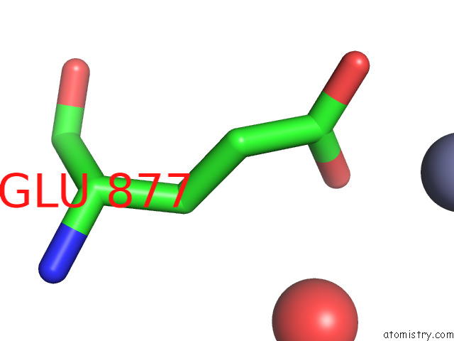
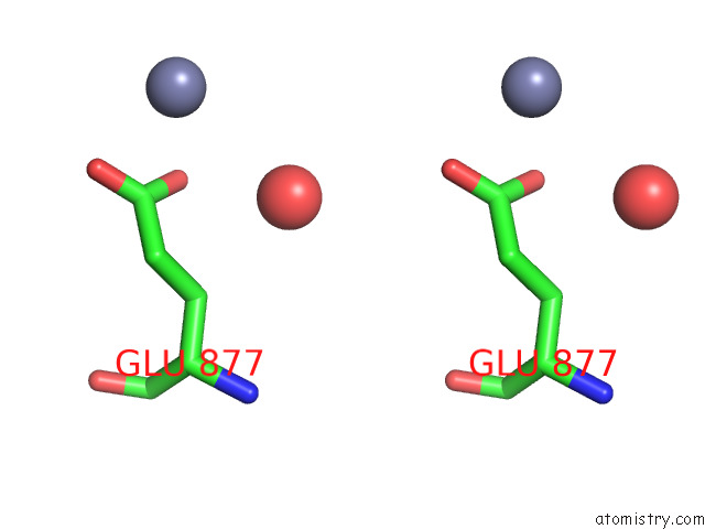
Zinc binding site 1 out of 4 in 7weg
Go back to
Zinc binding site 1 out
of 4 in the Complex Structure of PDZD7 and FCHSD2
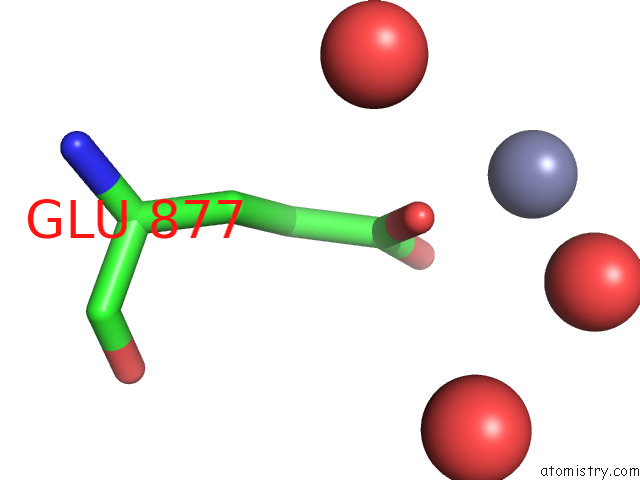
Mono view
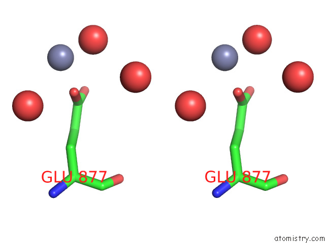
Stereo pair view
Mono view
Stereo pair view
A full contact list of Zinc with other atoms in the Zn binding
site number 1 of Complex Structure of PDZD7 and FCHSD2 within 5.0Å range:
|
Zinc binding site 2 out of 4 in 7weg
Go back to
Zinc binding site 2 out
of 4 in the Complex Structure of PDZD7 and FCHSD2
Mono view
Stereo pair view
Mono view
Stereo pair view
A full contact list of Zinc with other atoms in the Zn binding
site number 2 of Complex Structure of PDZD7 and FCHSD2 within 5.0Å range:
|
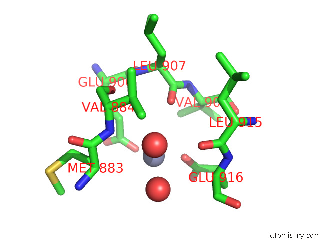
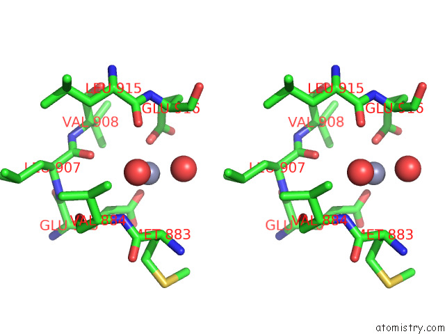
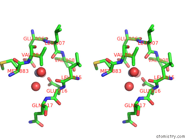
Zinc binding site 3 out of 4 in 7weg
Go back to
Zinc binding site 3 out
of 4 in the Complex Structure of PDZD7 and FCHSD2
Mono view
Stereo pair view
Mono view
Stereo pair view
A full contact list of Zinc with other atoms in the Zn binding
site number 3 of Complex Structure of PDZD7 and FCHSD2 within 5.0Å range:
|
Zinc binding site 4 out of 4 in 7weg
Go back to
Zinc binding site 4 out
of 4 in the Complex Structure of PDZD7 and FCHSD2
Mono view
Stereo pair view
Mono view
Stereo pair view
A full contact list of Zinc with other atoms in the Zn binding
site number 4 of Complex Structure of PDZD7 and FCHSD2 within 5.0Å range:
|
Reference:
H.Wang,
D.Zhao,
H.Du,
X.Zhai,
S.Wu,
L.Lin,
Z.Xu,
Q.Lu.
Deafness-Related Protein PDZD7 Forms Complex with the C-Terminal Tail of FCHSD2. Biochem.J. V. 479 1393 2022.
ISSN: ESSN 1470-8728
PubMed: 35695292
DOI: 10.1042/BCJ20220147
Page generated: Wed Oct 30 14:21:32 2024
ISSN: ESSN 1470-8728
PubMed: 35695292
DOI: 10.1042/BCJ20220147
Last articles
Zn in 9MJ5Zn in 9HNW
Zn in 9G0L
Zn in 9FNE
Zn in 9DZN
Zn in 9E0I
Zn in 9D32
Zn in 9DAK
Zn in 8ZXC
Zn in 8ZUF