Zinc in PDB 7r70: Crystal Structure of the UBARK2C Fusion Protein
Enzymatic activity of Crystal Structure of the UBARK2C Fusion Protein
All present enzymatic activity of Crystal Structure of the UBARK2C Fusion Protein:
2.3.2.27;
2.3.2.27;
Protein crystallography data
The structure of Crystal Structure of the UBARK2C Fusion Protein, PDB code: 7r70
was solved by
A.Paluda,
A.J.Middleton,
P.D.Mace,
C.L.Day,
with X-Ray Crystallography technique. A brief refinement statistics is given in the table below:
| Resolution Low / High (Å) | 44.59 / 2.50 |
| Space group | P 62 2 2 |
| Cell size a, b, c (Å), α, β, γ (°) | 154.47, 154.47, 81.706, 90, 90, 120 |
| R / Rfree (%) | 20 / 24.5 |
Zinc Binding Sites:
The binding sites of Zinc atom in the Crystal Structure of the UBARK2C Fusion Protein
(pdb code 7r70). This binding sites where shown within
5.0 Angstroms radius around Zinc atom.
In total 4 binding sites of Zinc where determined in the Crystal Structure of the UBARK2C Fusion Protein, PDB code: 7r70:
Jump to Zinc binding site number: 1; 2; 3; 4;
In total 4 binding sites of Zinc where determined in the Crystal Structure of the UBARK2C Fusion Protein, PDB code: 7r70:
Jump to Zinc binding site number: 1; 2; 3; 4;
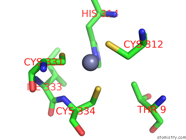
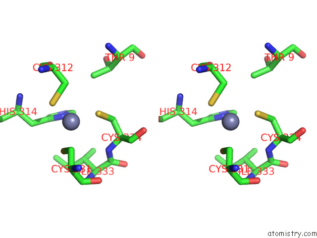
Zinc binding site 1 out of 4 in 7r70
Go back to
Zinc binding site 1 out
of 4 in the Crystal Structure of the UBARK2C Fusion Protein
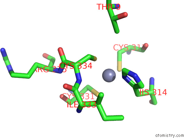
Mono view
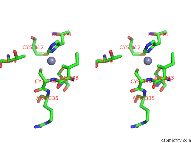
Stereo pair view
Mono view
Stereo pair view
A full contact list of Zinc with other atoms in the Zn binding
site number 1 of Crystal Structure of the UBARK2C Fusion Protein within 5.0Å range:
|
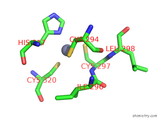
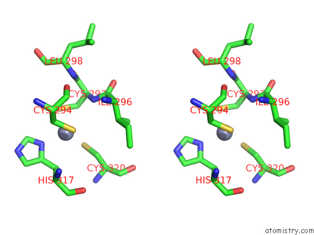
Zinc binding site 2 out of 4 in 7r70
Go back to
Zinc binding site 2 out
of 4 in the Crystal Structure of the UBARK2C Fusion Protein
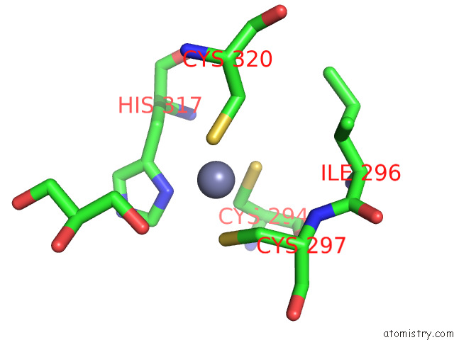
Mono view
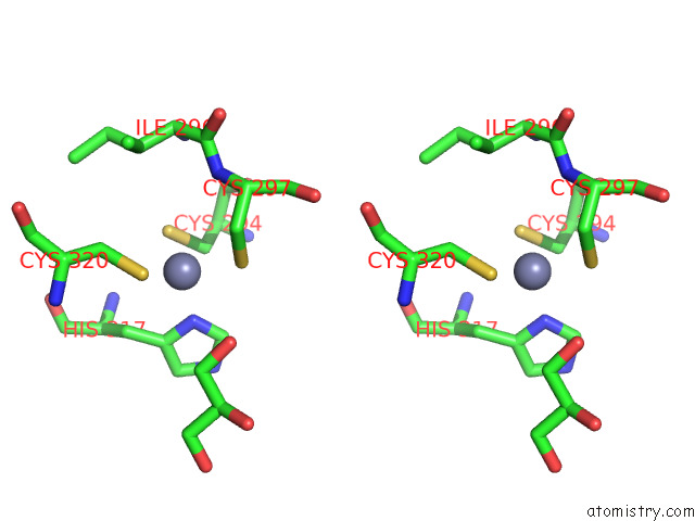
Stereo pair view
Mono view
Stereo pair view
A full contact list of Zinc with other atoms in the Zn binding
site number 2 of Crystal Structure of the UBARK2C Fusion Protein within 5.0Å range:
|
Zinc binding site 3 out of 4 in 7r70
Go back to
Zinc binding site 3 out
of 4 in the Crystal Structure of the UBARK2C Fusion Protein
Mono view
Stereo pair view
Mono view
Stereo pair view
A full contact list of Zinc with other atoms in the Zn binding
site number 3 of Crystal Structure of the UBARK2C Fusion Protein within 5.0Å range:
|
Zinc binding site 4 out of 4 in 7r70
Go back to
Zinc binding site 4 out
of 4 in the Crystal Structure of the UBARK2C Fusion Protein
Mono view
Stereo pair view
Mono view
Stereo pair view
A full contact list of Zinc with other atoms in the Zn binding
site number 4 of Crystal Structure of the UBARK2C Fusion Protein within 5.0Å range:
|
Reference:
A.Paluda,
A.J.Middleton,
C.Rossig,
P.D.Mace,
C.L.Day.
Ubiquitin and A Charged Loop Regulate the Ubiquitin E3 Ligase Activity of ARK2C. Nat Commun V. 13 1181 2022.
ISSN: ESSN 2041-1723
PubMed: 35246518
DOI: 10.1038/S41467-022-28782-Y
Page generated: Wed Oct 30 10:09:12 2024
ISSN: ESSN 2041-1723
PubMed: 35246518
DOI: 10.1038/S41467-022-28782-Y
Last articles
Zn in 9MJ5Zn in 9HNW
Zn in 9G0L
Zn in 9FNE
Zn in 9DZN
Zn in 9E0I
Zn in 9D32
Zn in 9DAK
Zn in 8ZXC
Zn in 8ZUF