Zinc in PDB 7qy5: Crystal Structure of the S.Pombe ARS2-RED1 Complex.
Protein crystallography data
The structure of Crystal Structure of the S.Pombe ARS2-RED1 Complex., PDB code: 7qy5
was solved by
A.E.Foucher,
J.Kadlec,
with X-Ray Crystallography technique. A brief refinement statistics is given in the table below:
| Resolution Low / High (Å) | 84.85 / 2.77 |
| Space group | P 1 21 1 |
| Cell size a, b, c (Å), α, β, γ (°) | 64.67, 128.281, 89.261, 90, 108.08, 90 |
| R / Rfree (%) | 22.8 / 29.8 |
Zinc Binding Sites:
The binding sites of Zinc atom in the Crystal Structure of the S.Pombe ARS2-RED1 Complex.
(pdb code 7qy5). This binding sites where shown within
5.0 Angstroms radius around Zinc atom.
In total 2 binding sites of Zinc where determined in the Crystal Structure of the S.Pombe ARS2-RED1 Complex., PDB code: 7qy5:
Jump to Zinc binding site number: 1; 2;
In total 2 binding sites of Zinc where determined in the Crystal Structure of the S.Pombe ARS2-RED1 Complex., PDB code: 7qy5:
Jump to Zinc binding site number: 1; 2;
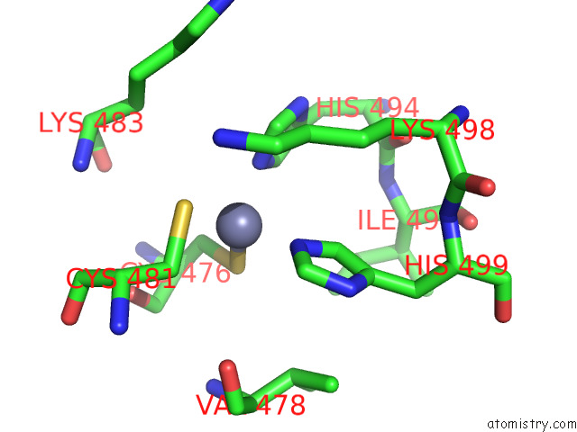
Zinc binding site 1 out of 2 in 7qy5
Go back to
Zinc binding site 1 out
of 2 in the Crystal Structure of the S.Pombe ARS2-RED1 Complex.
Mono view

Stereo pair view
Mono view
Stereo pair view
A full contact list of Zinc with other atoms in the Zn binding
site number 1 of Crystal Structure of the S.Pombe ARS2-RED1 Complex. within 5.0Å range:
|
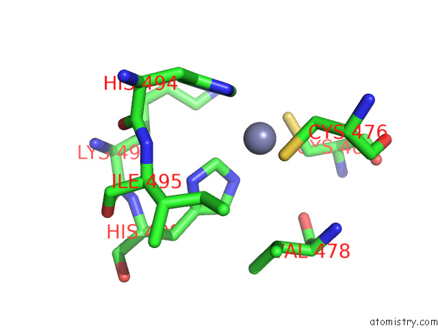
Zinc binding site 2 out of 2 in 7qy5
Go back to
Zinc binding site 2 out
of 2 in the Crystal Structure of the S.Pombe ARS2-RED1 Complex.
Mono view

Stereo pair view
Mono view
Stereo pair view
A full contact list of Zinc with other atoms in the Zn binding
site number 2 of Crystal Structure of the S.Pombe ARS2-RED1 Complex. within 5.0Å range:
|
Reference:
A.E.Foucher,
L.Touat-Todeschini,
A.B.Juarez-Martinez,
A.Rakitch,
H.Laroussi,
C.Karczewski,
S.Acajjaoui,
M.Soler-Lopez,
S.Cusack,
C.D.Mackereth,
A.Verdel,
J.Kadlec.
Structural Analysis of RED1 As A Conserved Scaffold of the Rna-Targeting Mtrec/Paxt Complex. Nat Commun V. 13 4969 2022.
ISSN: ESSN 2041-1723
PubMed: 36002457
DOI: 10.1038/S41467-022-32542-3
Page generated: Wed Oct 30 10:03:24 2024
ISSN: ESSN 2041-1723
PubMed: 36002457
DOI: 10.1038/S41467-022-32542-3
Last articles
Zn in 9MJ5Zn in 9HNW
Zn in 9G0L
Zn in 9FNE
Zn in 9DZN
Zn in 9E0I
Zn in 9D32
Zn in 9DAK
Zn in 8ZXC
Zn in 8ZUF