Zinc in PDB 7p2k: Solution uc(Nmr) Structure of Arginine to Cysteine Mutant of Arkadia Ring Domain.
Enzymatic activity of Solution uc(Nmr) Structure of Arginine to Cysteine Mutant of Arkadia Ring Domain.
All present enzymatic activity of Solution uc(Nmr) Structure of Arginine to Cysteine Mutant of Arkadia Ring Domain.:
2.3.2.27;
2.3.2.27;
Zinc Binding Sites:
The binding sites of Zinc atom in the Solution uc(Nmr) Structure of Arginine to Cysteine Mutant of Arkadia Ring Domain.
(pdb code 7p2k). This binding sites where shown within
5.0 Angstroms radius around Zinc atom.
In total 2 binding sites of Zinc where determined in the Solution uc(Nmr) Structure of Arginine to Cysteine Mutant of Arkadia Ring Domain., PDB code: 7p2k:
Jump to Zinc binding site number: 1; 2;
In total 2 binding sites of Zinc where determined in the Solution uc(Nmr) Structure of Arginine to Cysteine Mutant of Arkadia Ring Domain., PDB code: 7p2k:
Jump to Zinc binding site number: 1; 2;
Zinc binding site 1 out of 2 in 7p2k
Go back to
Zinc binding site 1 out
of 2 in the Solution uc(Nmr) Structure of Arginine to Cysteine Mutant of Arkadia Ring Domain.
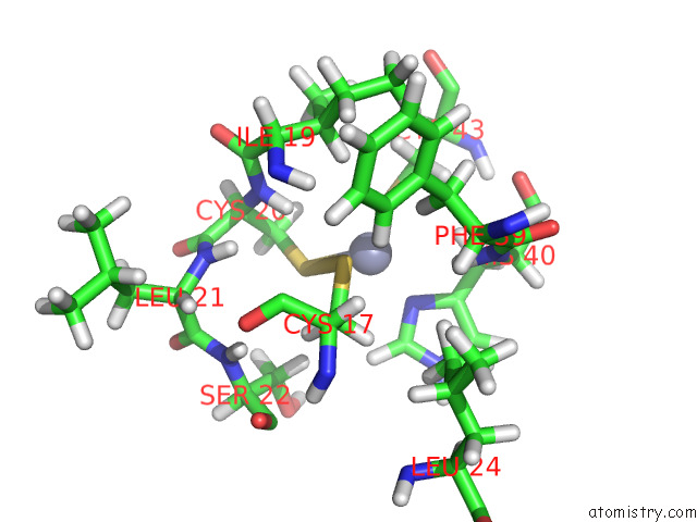
Mono view
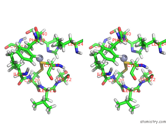
Stereo pair view
Mono view
Stereo pair view
A full contact list of Zinc with other atoms in the Zn binding
site number 1 of Solution uc(Nmr) Structure of Arginine to Cysteine Mutant of Arkadia Ring Domain. within 5.0Å range:
|
Zinc binding site 2 out of 2 in 7p2k
Go back to
Zinc binding site 2 out
of 2 in the Solution uc(Nmr) Structure of Arginine to Cysteine Mutant of Arkadia Ring Domain.
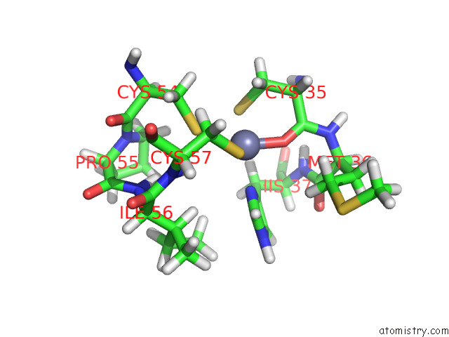
Mono view
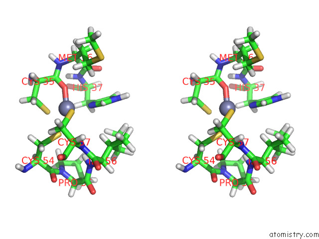
Stereo pair view
Mono view
Stereo pair view
A full contact list of Zinc with other atoms in the Zn binding
site number 2 of Solution uc(Nmr) Structure of Arginine to Cysteine Mutant of Arkadia Ring Domain. within 5.0Å range:
|
Reference:
M.Birkou,
V.Raptis,
K.D.Marousis,
A.Tsevis,
K.Bourikas,
D.Bentrop,
V.Episkopou,
G.A.Spyroulias.
Impact of A Single Nucleotide Polymorphism on the 3D Protein Structure and Ubiquitination Activity of E3 Ubiquitin Ligase Arkadia. Front Mol Biosci V. 9 44129 2022.
ISSN: ESSN 2296-889X
PubMed: 35281275
DOI: 10.3389/FMOLB.2022.844129
Page generated: Wed Oct 30 08:58:14 2024
ISSN: ESSN 2296-889X
PubMed: 35281275
DOI: 10.3389/FMOLB.2022.844129
Last articles
Zn in 9MJ5Zn in 9HNW
Zn in 9G0L
Zn in 9FNE
Zn in 9DZN
Zn in 9E0I
Zn in 9D32
Zn in 9DAK
Zn in 8ZXC
Zn in 8ZUF