Zinc in PDB 7n1s: Crystal Structure Analysis of Xac Nucleotide Pyrophosphatase/Phosphodiesterase
Protein crystallography data
The structure of Crystal Structure Analysis of Xac Nucleotide Pyrophosphatase/Phosphodiesterase, PDB code: 7n1s
was solved by
D.Fernandez,
L.Li,
J.A.Brown,
J.A.Carozza,
with X-Ray Crystallography technique. A brief refinement statistics is given in the table below:
| Resolution Low / High (Å) | 30.61 / 2.00 |
| Space group | P 21 21 21 |
| Cell size a, b, c (Å), α, β, γ (°) | 65.204, 77.712, 129.292, 90, 90, 90 |
| R / Rfree (%) | 21.4 / 25.7 |
Zinc Binding Sites:
The binding sites of Zinc atom in the Crystal Structure Analysis of Xac Nucleotide Pyrophosphatase/Phosphodiesterase
(pdb code 7n1s). This binding sites where shown within
5.0 Angstroms radius around Zinc atom.
In total 2 binding sites of Zinc where determined in the Crystal Structure Analysis of Xac Nucleotide Pyrophosphatase/Phosphodiesterase, PDB code: 7n1s:
Jump to Zinc binding site number: 1; 2;
In total 2 binding sites of Zinc where determined in the Crystal Structure Analysis of Xac Nucleotide Pyrophosphatase/Phosphodiesterase, PDB code: 7n1s:
Jump to Zinc binding site number: 1; 2;
Zinc binding site 1 out of 2 in 7n1s
Go back to
Zinc binding site 1 out
of 2 in the Crystal Structure Analysis of Xac Nucleotide Pyrophosphatase/Phosphodiesterase
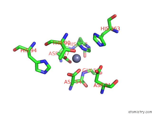
Mono view
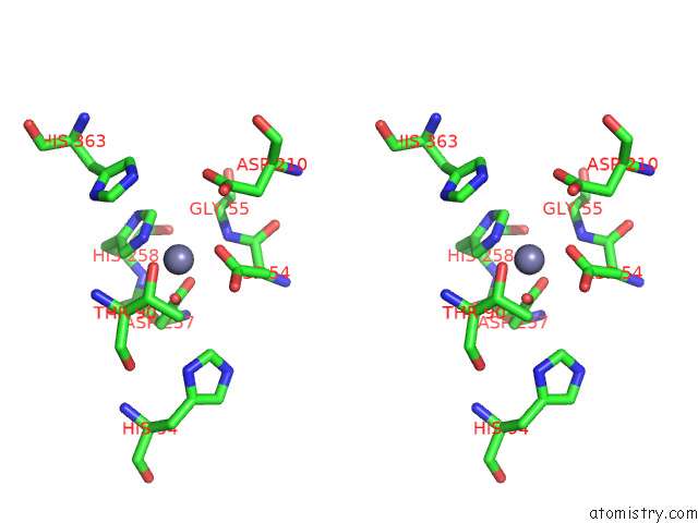
Stereo pair view
Mono view
Stereo pair view
A full contact list of Zinc with other atoms in the Zn binding
site number 1 of Crystal Structure Analysis of Xac Nucleotide Pyrophosphatase/Phosphodiesterase within 5.0Å range:
|
Zinc binding site 2 out of 2 in 7n1s
Go back to
Zinc binding site 2 out
of 2 in the Crystal Structure Analysis of Xac Nucleotide Pyrophosphatase/Phosphodiesterase
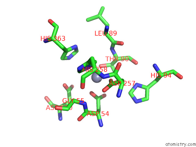
Mono view
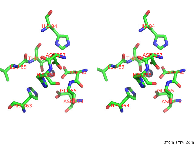
Stereo pair view
Mono view
Stereo pair view
A full contact list of Zinc with other atoms in the Zn binding
site number 2 of Crystal Structure Analysis of Xac Nucleotide Pyrophosphatase/Phosphodiesterase within 5.0Å range:
|
Reference:
J.A.Carozza,
A.F.Cordova,
J.A.Brown,
Y.Alsaif,
V.Bohnert,
X.Cao,
R.E.Mardjuki,
G.Skariah,
D.Fernandez,
L.Li.
ENPP1'S Regulation of Extracellular Cgamp Is A Ubiquitous Mechanism of Attenuating Sting Signaling. Proc.Natl.Acad.Sci.Usa V. 119 89119 2022.
ISSN: ESSN 1091-6490
PubMed: 35588451
DOI: 10.1073/PNAS.2119189119
Page generated: Wed Oct 30 07:38:40 2024
ISSN: ESSN 1091-6490
PubMed: 35588451
DOI: 10.1073/PNAS.2119189119
Last articles
Zn in 9MJ5Zn in 9HNW
Zn in 9G0L
Zn in 9FNE
Zn in 9DZN
Zn in 9E0I
Zn in 9D32
Zn in 9DAK
Zn in 8ZXC
Zn in 8ZUF