Zinc in PDB 7mo4: Crystal Structure of the ZNF3 of Nucleoporin NUP153 in Complex with Ran-Gdp, Resolution 2.4 Angstrom
Protein crystallography data
The structure of Crystal Structure of the ZNF3 of Nucleoporin NUP153 in Complex with Ran-Gdp, Resolution 2.4 Angstrom, PDB code: 7mo4
was solved by
C.J.Bley,
S.Nie,
G.W.Mobbs,
S.Petrovic,
A.T.Gres,
X.Liu,
S.Mukherjee,
S.Harvey,
F.M.Huber,
D.H.Lin,
B.Brown,
A.W.Tang,
E.J.Rundlet,
A.R.Correia,
S.Chen,
S.G.Regmi,
T.A.Stevens,
C.A.Jette,
M.Dasso,
A.Patke,
A.F.Palazzo,
A.A.Kossiakoff,
A.Hoelz,
with X-Ray Crystallography technique. A brief refinement statistics is given in the table below:
| Resolution Low / High (Å) | 29.44 / 2.40 |
| Space group | P 1 21 1 |
| Cell size a, b, c (Å), α, β, γ (°) | 67.66, 61.24, 69.75, 90, 104.04, 90 |
| R / Rfree (%) | 21.4 / 23.7 |
Other elements in 7mo4:
The structure of Crystal Structure of the ZNF3 of Nucleoporin NUP153 in Complex with Ran-Gdp, Resolution 2.4 Angstrom also contains other interesting chemical elements:
| Magnesium | (Mg) | 2 atoms |
Zinc Binding Sites:
The binding sites of Zinc atom in the Crystal Structure of the ZNF3 of Nucleoporin NUP153 in Complex with Ran-Gdp, Resolution 2.4 Angstrom
(pdb code 7mo4). This binding sites where shown within
5.0 Angstroms radius around Zinc atom.
In total 2 binding sites of Zinc where determined in the Crystal Structure of the ZNF3 of Nucleoporin NUP153 in Complex with Ran-Gdp, Resolution 2.4 Angstrom, PDB code: 7mo4:
Jump to Zinc binding site number: 1; 2;
In total 2 binding sites of Zinc where determined in the Crystal Structure of the ZNF3 of Nucleoporin NUP153 in Complex with Ran-Gdp, Resolution 2.4 Angstrom, PDB code: 7mo4:
Jump to Zinc binding site number: 1; 2;
Zinc binding site 1 out of 2 in 7mo4
Go back to
Zinc binding site 1 out
of 2 in the Crystal Structure of the ZNF3 of Nucleoporin NUP153 in Complex with Ran-Gdp, Resolution 2.4 Angstrom
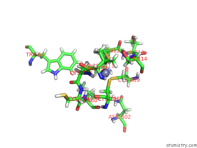
Mono view
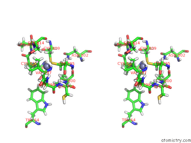
Stereo pair view
Mono view
Stereo pair view
A full contact list of Zinc with other atoms in the Zn binding
site number 1 of Crystal Structure of the ZNF3 of Nucleoporin NUP153 in Complex with Ran-Gdp, Resolution 2.4 Angstrom within 5.0Å range:
|
Zinc binding site 2 out of 2 in 7mo4
Go back to
Zinc binding site 2 out
of 2 in the Crystal Structure of the ZNF3 of Nucleoporin NUP153 in Complex with Ran-Gdp, Resolution 2.4 Angstrom
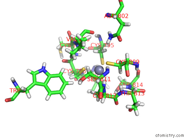
Mono view
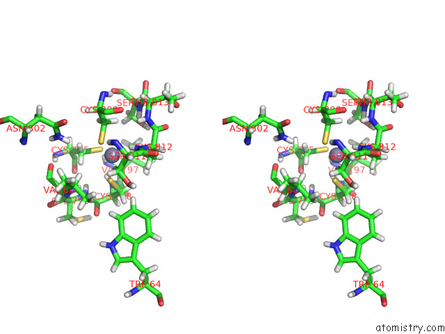
Stereo pair view
Mono view
Stereo pair view
A full contact list of Zinc with other atoms in the Zn binding
site number 2 of Crystal Structure of the ZNF3 of Nucleoporin NUP153 in Complex with Ran-Gdp, Resolution 2.4 Angstrom within 5.0Å range:
|
Reference:
C.J.Bley,
S.Nie,
G.W.Mobbs,
S.Petrovic,
A.T.Gres,
X.Liu,
S.Mukherjee,
S.Harvey,
F.M.Huber,
D.H.Lin,
B.Brown,
A.W.Tang,
E.J.Rundlet,
A.R.Correia,
S.Chen,
S.G.Regmi,
T.A.Stevens,
C.A.Jette,
M.Dasso,
A.Patke,
A.F.Palazzo,
A.A.Kossiakoff,
A.Hoelz.
Architecture of the Cytoplasmic Face of the Nuclear Pore. Science V. 376 M9129 2022.
ISSN: ESSN 1095-9203
PubMed: 35679405
DOI: 10.1126/SCIENCE.ABM9129
Page generated: Wed Oct 30 07:34:10 2024
ISSN: ESSN 1095-9203
PubMed: 35679405
DOI: 10.1126/SCIENCE.ABM9129
Last articles
Zn in 9MJ5Zn in 9HNW
Zn in 9G0L
Zn in 9FNE
Zn in 9DZN
Zn in 9E0I
Zn in 9D32
Zn in 9DAK
Zn in 8ZXC
Zn in 8ZUF