Zinc in PDB 7kyh: Botulism Neurooxin Light Chain A App Form
Protein crystallography data
The structure of Botulism Neurooxin Light Chain A App Form, PDB code: 7kyh
was solved by
M.E.Ortega,
N.T.Salzameda,
with X-Ray Crystallography technique. A brief refinement statistics is given in the table below:
| Resolution Low / High (Å) | 48.09 / 2.91 |
| Space group | P 1 |
| Cell size a, b, c (Å), α, β, γ (°) | 57.315, 84.05, 99.723, 103.78, 91.84, 108.67 |
| R / Rfree (%) | 13.8 / 17.3 |
Other elements in 7kyh:
The structure of Botulism Neurooxin Light Chain A App Form also contains other interesting chemical elements:
| Chlorine | (Cl) | 8 atoms |
Zinc Binding Sites:
The binding sites of Zinc atom in the Botulism Neurooxin Light Chain A App Form
(pdb code 7kyh). This binding sites where shown within
5.0 Angstroms radius around Zinc atom.
In total 4 binding sites of Zinc where determined in the Botulism Neurooxin Light Chain A App Form, PDB code: 7kyh:
Jump to Zinc binding site number: 1; 2; 3; 4;
In total 4 binding sites of Zinc where determined in the Botulism Neurooxin Light Chain A App Form, PDB code: 7kyh:
Jump to Zinc binding site number: 1; 2; 3; 4;
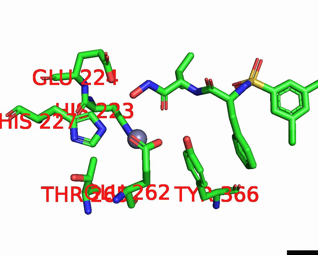
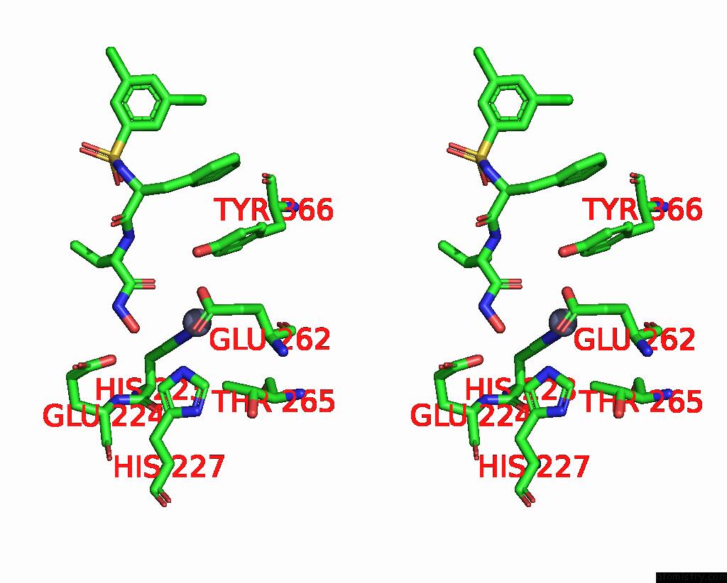
Zinc binding site 1 out of 4 in 7kyh
Go back to
Zinc binding site 1 out
of 4 in the Botulism Neurooxin Light Chain A App Form
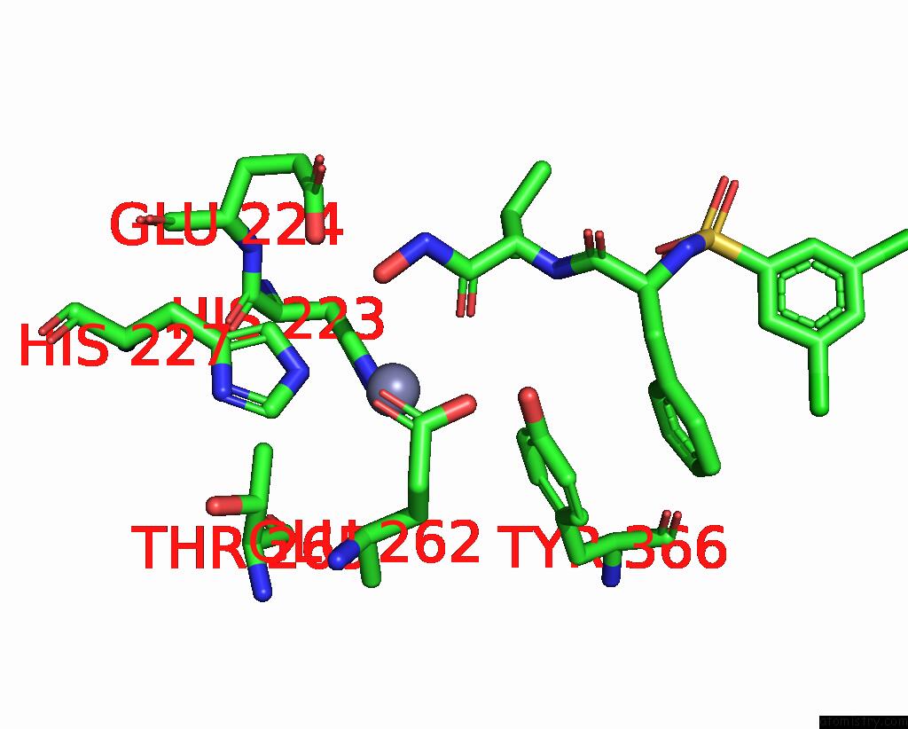
Mono view
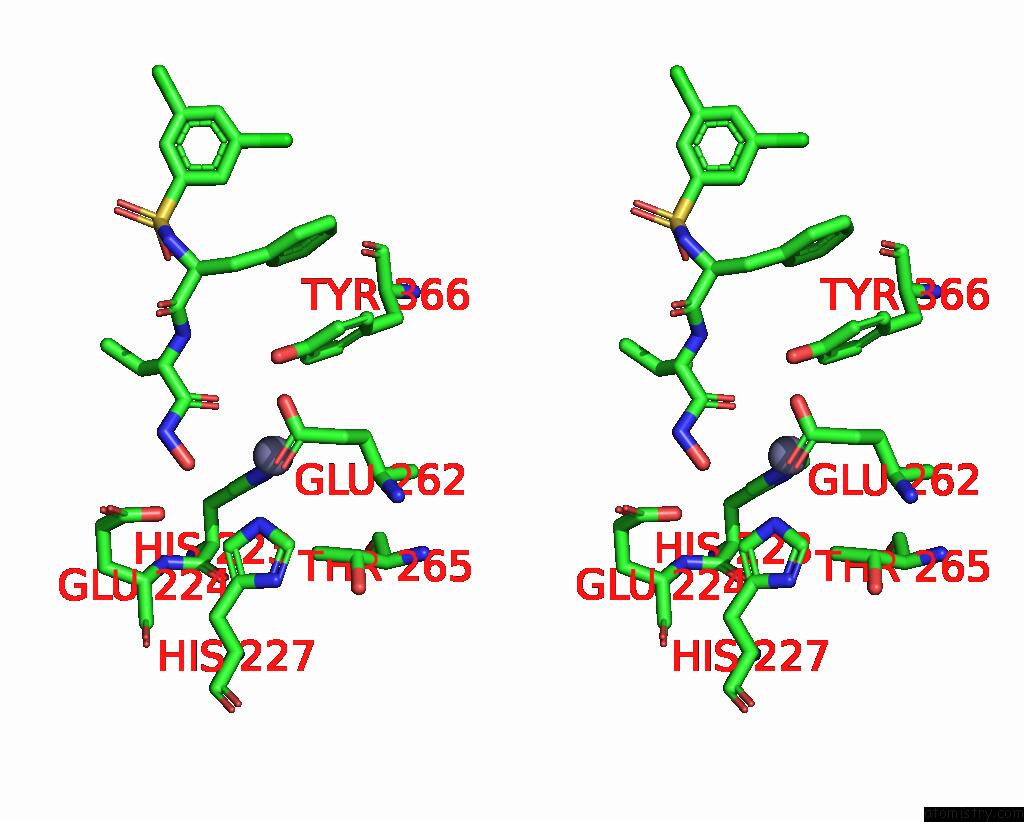
Stereo pair view
Mono view
Stereo pair view
A full contact list of Zinc with other atoms in the Zn binding
site number 1 of Botulism Neurooxin Light Chain A App Form within 5.0Å range:
|
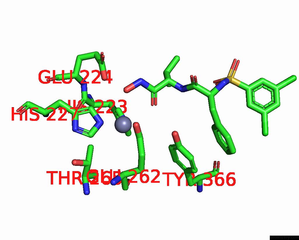
Zinc binding site 2 out of 4 in 7kyh
Go back to
Zinc binding site 2 out
of 4 in the Botulism Neurooxin Light Chain A App Form
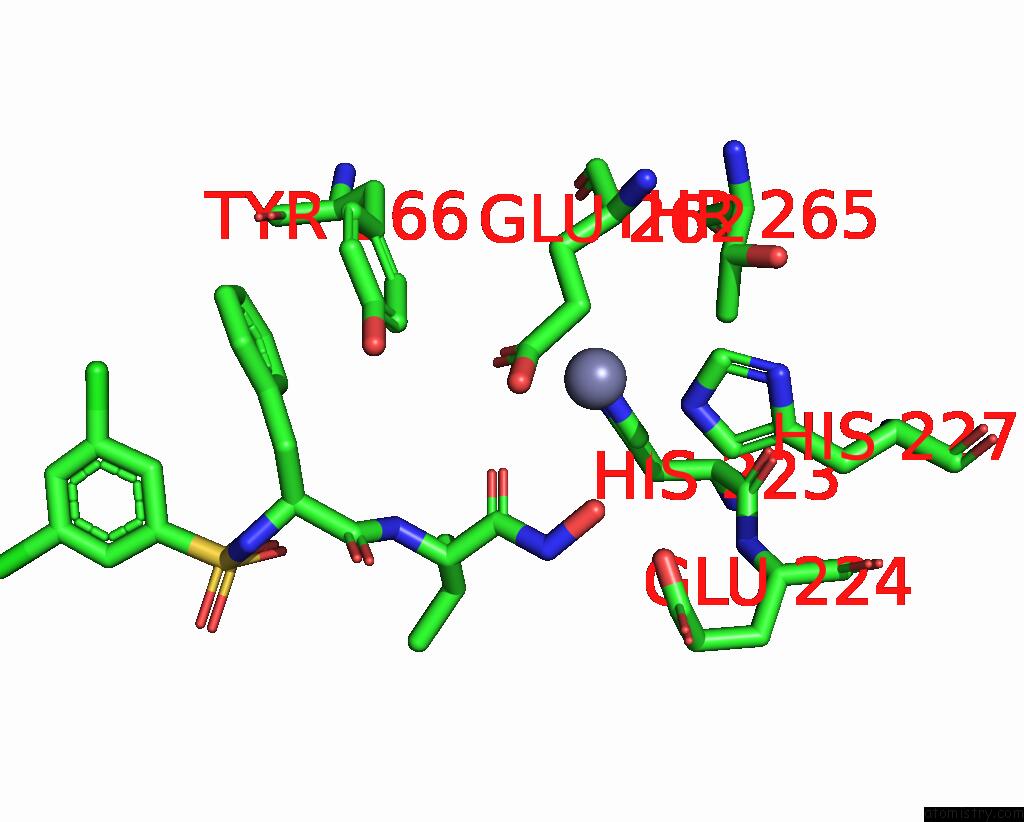
Mono view
Stereo pair view
Mono view
Stereo pair view
A full contact list of Zinc with other atoms in the Zn binding
site number 2 of Botulism Neurooxin Light Chain A App Form within 5.0Å range:
|
Zinc binding site 3 out of 4 in 7kyh
Go back to
Zinc binding site 3 out
of 4 in the Botulism Neurooxin Light Chain A App Form
Mono view
Stereo pair view
Mono view
Stereo pair view
A full contact list of Zinc with other atoms in the Zn binding
site number 3 of Botulism Neurooxin Light Chain A App Form within 5.0Å range:
|
Zinc binding site 4 out of 4 in 7kyh
Go back to
Zinc binding site 4 out
of 4 in the Botulism Neurooxin Light Chain A App Form
Mono view
Stereo pair view
Mono view
Stereo pair view
A full contact list of Zinc with other atoms in the Zn binding
site number 4 of Botulism Neurooxin Light Chain A App Form within 5.0Å range:
|
Reference:
M.Amezcua,
R.S.Cruz,
A.Ku,
W.Moran,
M.E.Ortega,
N.T.Salzameda.
Discovery of Dipeptides As Potent Botulinum Neurotoxin A Light-Chain Inhibitors. Acs Med.Chem.Lett. V. 12 295 2021.
ISSN: ISSN 1948-5875
PubMed: 33603978
DOI: 10.1021/ACSMEDCHEMLETT.0C00674
Page generated: Tue Oct 29 22:33:44 2024
ISSN: ISSN 1948-5875
PubMed: 33603978
DOI: 10.1021/ACSMEDCHEMLETT.0C00674
Last articles
Zn in 9MJ5Zn in 9HNW
Zn in 9G0L
Zn in 9FNE
Zn in 9DZN
Zn in 9E0I
Zn in 9D32
Zn in 9DAK
Zn in 8ZXC
Zn in 8ZUF