Zinc in PDB 7k95: Crystal Structure of Human CPSF30 in Complex with HFIP1
Protein crystallography data
The structure of Crystal Structure of Human CPSF30 in Complex with HFIP1, PDB code: 7k95
was solved by
K.Hamilton,
L.Tong,
with X-Ray Crystallography technique. A brief refinement statistics is given in the table below:
| Resolution Low / High (Å) | 39.55 / 1.90 |
| Space group | P 64 |
| Cell size a, b, c (Å), α, β, γ (°) | 79.040, 79.040, 48.662, 90.00, 90.00, 120.00 |
| R / Rfree (%) | 17.9 / 20.9 |
Zinc Binding Sites:
The binding sites of Zinc atom in the Crystal Structure of Human CPSF30 in Complex with HFIP1
(pdb code 7k95). This binding sites where shown within
5.0 Angstroms radius around Zinc atom.
In total 2 binding sites of Zinc where determined in the Crystal Structure of Human CPSF30 in Complex with HFIP1, PDB code: 7k95:
Jump to Zinc binding site number: 1; 2;
In total 2 binding sites of Zinc where determined in the Crystal Structure of Human CPSF30 in Complex with HFIP1, PDB code: 7k95:
Jump to Zinc binding site number: 1; 2;
Zinc binding site 1 out of 2 in 7k95
Go back to
Zinc binding site 1 out
of 2 in the Crystal Structure of Human CPSF30 in Complex with HFIP1
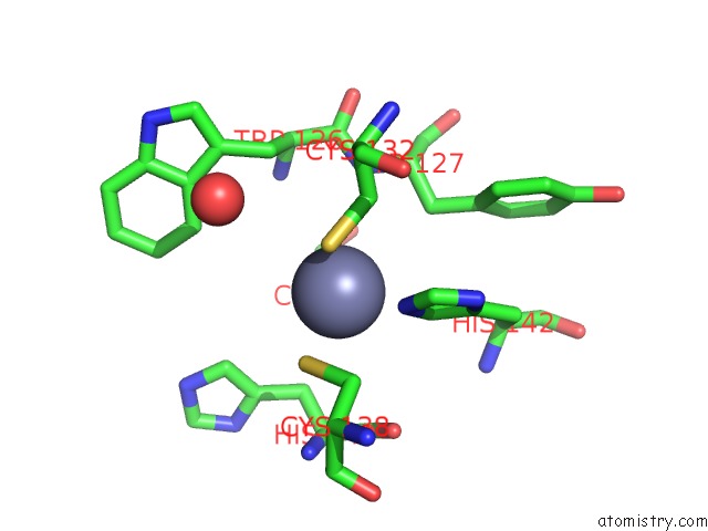
Mono view
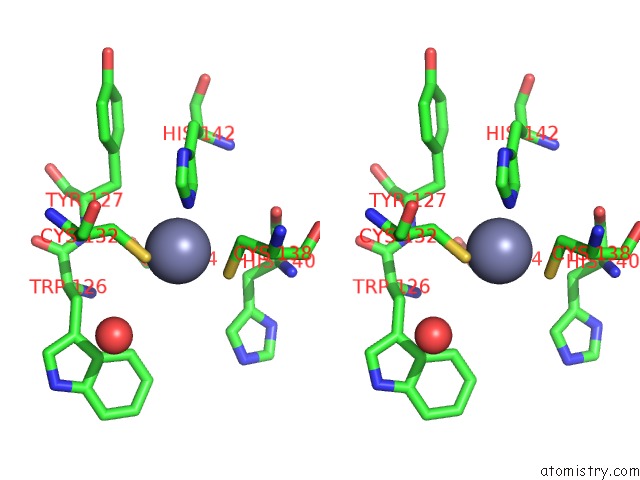
Stereo pair view
Mono view
Stereo pair view
A full contact list of Zinc with other atoms in the Zn binding
site number 1 of Crystal Structure of Human CPSF30 in Complex with HFIP1 within 5.0Å range:
|
Zinc binding site 2 out of 2 in 7k95
Go back to
Zinc binding site 2 out
of 2 in the Crystal Structure of Human CPSF30 in Complex with HFIP1
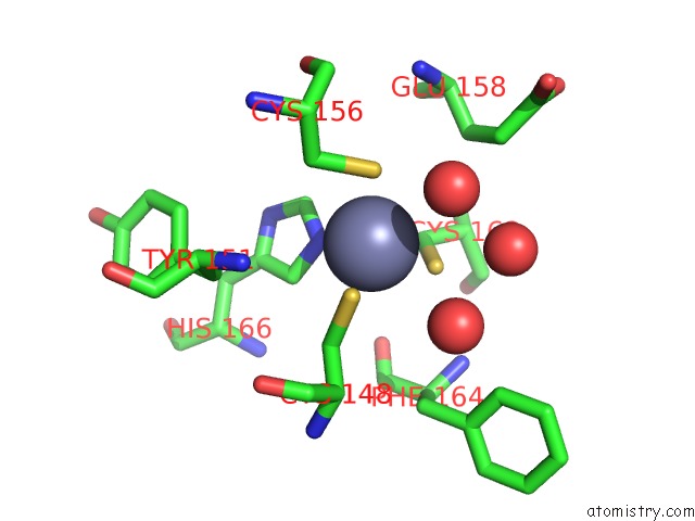
Mono view
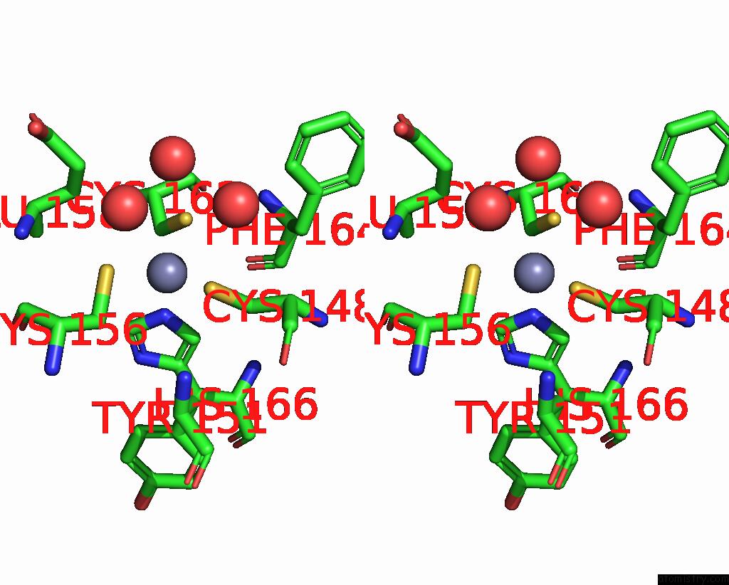
Stereo pair view
Mono view
Stereo pair view
A full contact list of Zinc with other atoms in the Zn binding
site number 2 of Crystal Structure of Human CPSF30 in Complex with HFIP1 within 5.0Å range:
|
Reference:
K.Hamilton,
L.Tong.
Molecular Mechanism For the Interaction Between Human CPSF30 and HFIP1. Genes Dev. 2020.
ISSN: ISSN 0890-9369
PubMed: 33122294
DOI: 10.1101/GAD.343814.120
Page generated: Tue Oct 29 21:19:29 2024
ISSN: ISSN 0890-9369
PubMed: 33122294
DOI: 10.1101/GAD.343814.120
Last articles
Zn in 9MJ5Zn in 9HNW
Zn in 9G0L
Zn in 9FNE
Zn in 9DZN
Zn in 9E0I
Zn in 9D32
Zn in 9DAK
Zn in 8ZXC
Zn in 8ZUF