Zinc »
PDB 7hl9-7jit »
7hof »
Zinc in PDB 7hof: Group Deposition For Crystallographic Fragment Screening of Coxsackievirus A16 (G-10) 2A Protease -- Crystal Structure of Coxsackievirus A16 (G-10) 2A Protease in Complex with Z1201620232 (A71EV2A-X0514)
Enzymatic activity of Group Deposition For Crystallographic Fragment Screening of Coxsackievirus A16 (G-10) 2A Protease -- Crystal Structure of Coxsackievirus A16 (G-10) 2A Protease in Complex with Z1201620232 (A71EV2A-X0514)
All present enzymatic activity of Group Deposition For Crystallographic Fragment Screening of Coxsackievirus A16 (G-10) 2A Protease -- Crystal Structure of Coxsackievirus A16 (G-10) 2A Protease in Complex with Z1201620232 (A71EV2A-X0514):
3.4.22.29;
3.4.22.29;
Protein crystallography data
The structure of Group Deposition For Crystallographic Fragment Screening of Coxsackievirus A16 (G-10) 2A Protease -- Crystal Structure of Coxsackievirus A16 (G-10) 2A Protease in Complex with Z1201620232 (A71EV2A-X0514), PDB code: 7hof
was solved by
R.M.Lithgo,
M.Fairhead,
L.Koekemoer,
B.H.Balcomb,
E.Capkin,
A.V.Chandran,
M.Golding,
A.S.Godoy,
J.C.Aschenbrenner,
P.G.Marples,
X.Ni,
W.Thompson,
C.W.E.Tomlinson,
C.Wild,
M.Winokan,
M.-A.E.Xavier,
D.Fearon,
F.Von Delft,
with X-Ray Crystallography technique. A brief refinement statistics is given in the table below:
| Resolution Low / High (Å) | 43.24 / 1.30 |
| Space group | C 1 2 1 |
| Cell size a, b, c (Å), α, β, γ (°) | 86.629, 56.969, 64.571, 90, 94.18, 90 |
| R / Rfree (%) | 20.6 / 23.7 |
Zinc Binding Sites:
The binding sites of Zinc atom in the Group Deposition For Crystallographic Fragment Screening of Coxsackievirus A16 (G-10) 2A Protease -- Crystal Structure of Coxsackievirus A16 (G-10) 2A Protease in Complex with Z1201620232 (A71EV2A-X0514)
(pdb code 7hof). This binding sites where shown within
5.0 Angstroms radius around Zinc atom.
In total 2 binding sites of Zinc where determined in the Group Deposition For Crystallographic Fragment Screening of Coxsackievirus A16 (G-10) 2A Protease -- Crystal Structure of Coxsackievirus A16 (G-10) 2A Protease in Complex with Z1201620232 (A71EV2A-X0514), PDB code: 7hof:
Jump to Zinc binding site number: 1; 2;
Zinc binding site 1 out of 2 in 7hof
Go back to
Zinc binding site 1 out
of 2 in the Group Deposition For Crystallographic Fragment Screening of Coxsackievirus A16 (G-10) 2A Protease -- Crystal Structure of Coxsackievirus A16 (G-10) 2A Protease in Complex with Z1201620232 (A71EV2A-X0514)
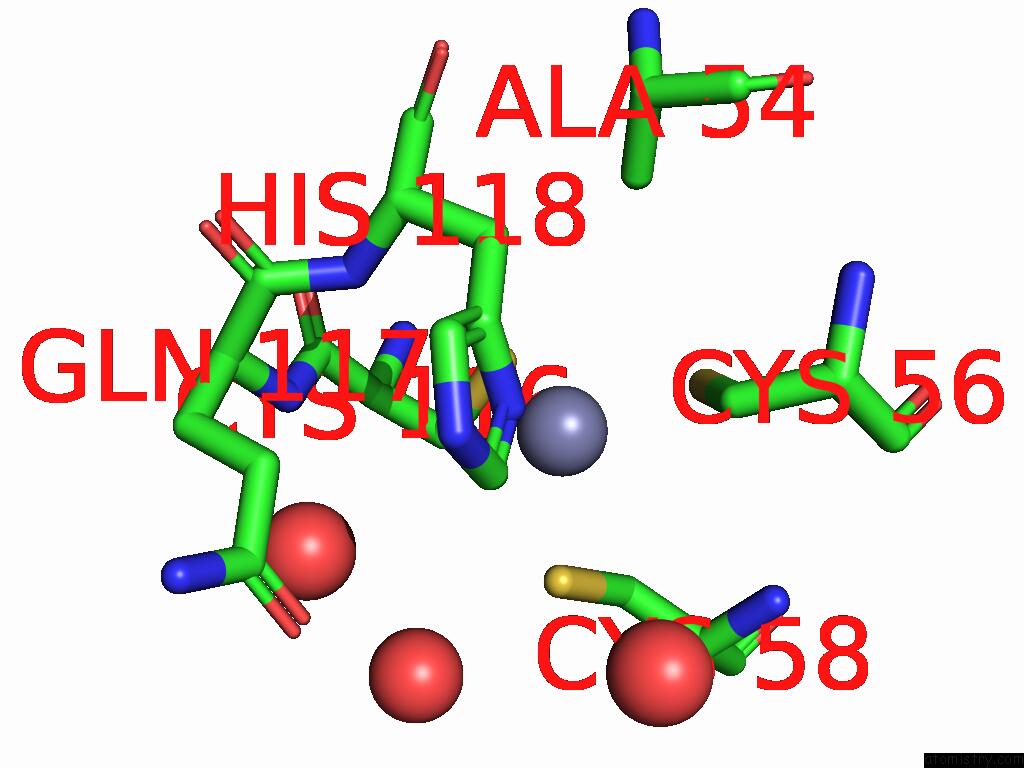
Mono view
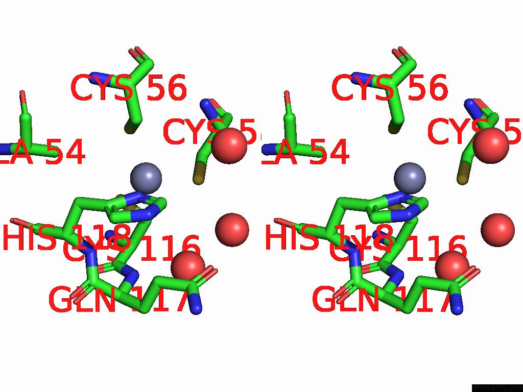
Stereo pair view
Mono view
Stereo pair view
A full contact list of Zinc with other atoms in the Zn binding
site number 1 of Group Deposition For Crystallographic Fragment Screening of Coxsackievirus A16 (G-10) 2A Protease -- Crystal Structure of Coxsackievirus A16 (G-10) 2A Protease in Complex with Z1201620232 (A71EV2A-X0514) within 5.0Å range:
|
Zinc binding site 2 out of 2 in 7hof
Go back to
Zinc binding site 2 out
of 2 in the Group Deposition For Crystallographic Fragment Screening of Coxsackievirus A16 (G-10) 2A Protease -- Crystal Structure of Coxsackievirus A16 (G-10) 2A Protease in Complex with Z1201620232 (A71EV2A-X0514)
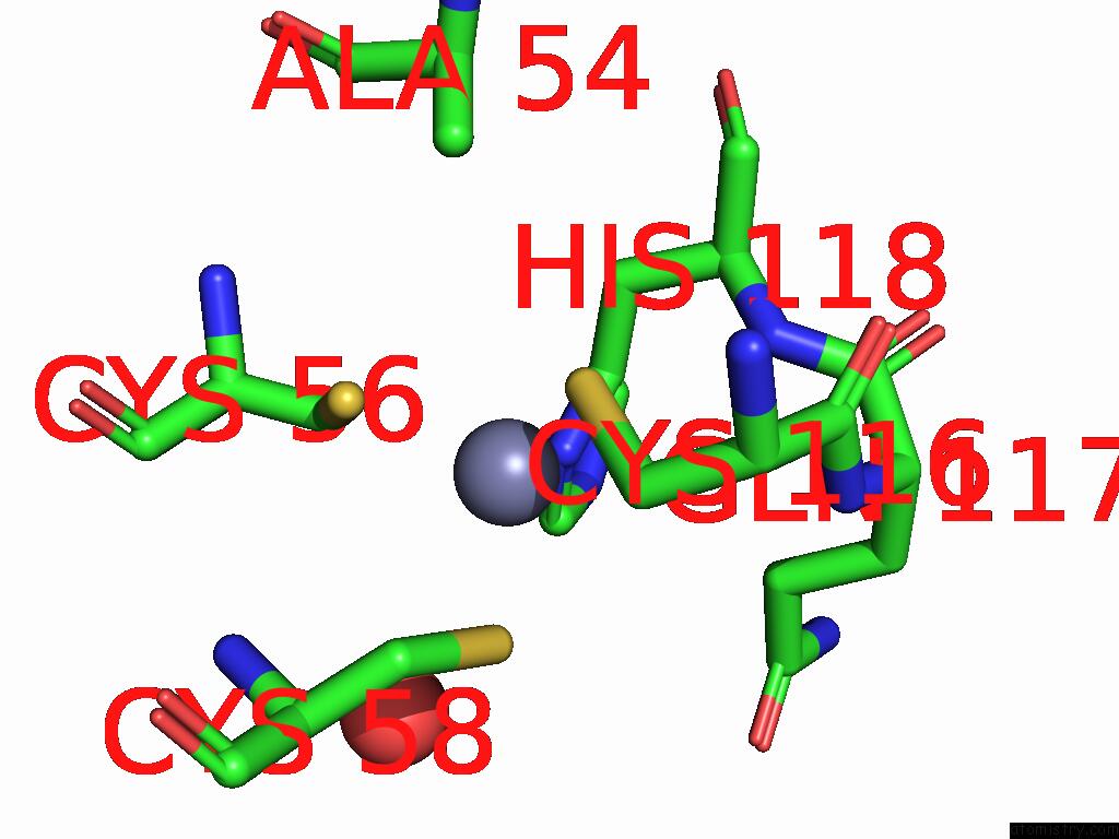
Mono view
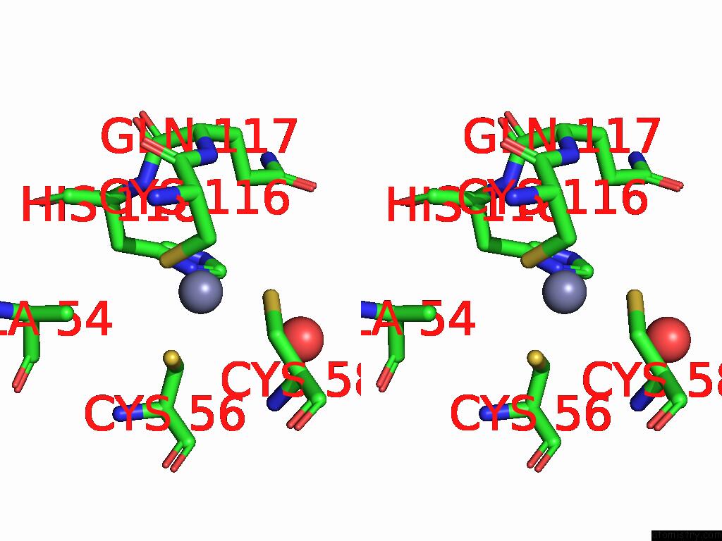
Stereo pair view
Mono view
Stereo pair view
A full contact list of Zinc with other atoms in the Zn binding
site number 2 of Group Deposition For Crystallographic Fragment Screening of Coxsackievirus A16 (G-10) 2A Protease -- Crystal Structure of Coxsackievirus A16 (G-10) 2A Protease in Complex with Z1201620232 (A71EV2A-X0514) within 5.0Å range:
|
Reference:
R.M.Lithgo,
M.Fairhead,
L.Koekemoer,
B.H.Balcomb,
E.Capkin,
A.V.Chandran,
M.Golding,
A.S.Godoy,
J.C.Aschenbrenner,
P.G.Marples,
X.Ni,
W.Thompson,
C.W.E.Tomlinson,
C.Wild,
M.Winokan,
M.-A.E.Xavier,
D.Fearon,
F.Von Delft.
Group Deposition For Crystallographic Fragment Screening of Coxsackievirus A16 (G-10) 2A Protease To Be Published.
Page generated: Fri Aug 22 00:45:37 2025
Last articles
Zn in 7YIVZn in 7YPB
Zn in 7YPA
Zn in 7YP9
Zn in 7YP8
Zn in 7YKA
Zn in 7YMO
Zn in 7YLQ
Zn in 7YHC
Zn in 7YIX