Zinc »
PDB 7f9d-7ftt »
7far »
Zinc in PDB 7far: Crystal Structure of PDE5A in Complex with Inhibitor L12
Enzymatic activity of Crystal Structure of PDE5A in Complex with Inhibitor L12
All present enzymatic activity of Crystal Structure of PDE5A in Complex with Inhibitor L12:
3.1.4.35;
3.1.4.35;
Protein crystallography data
The structure of Crystal Structure of PDE5A in Complex with Inhibitor L12, PDB code: 7far
was solved by
D.Wu,
Y.Y.Huang,
H.B.Luo,
with X-Ray Crystallography technique. A brief refinement statistics is given in the table below:
| Resolution Low / High (Å) | 23.80 / 2.40 |
| Space group | P 31 2 1 |
| Cell size a, b, c (Å), α, β, γ (°) | 73.917, 73.917, 132.251, 90, 90, 120 |
| R / Rfree (%) | 21.3 / 25 |
Other elements in 7far:
The structure of Crystal Structure of PDE5A in Complex with Inhibitor L12 also contains other interesting chemical elements:
| Fluorine | (F) | 2 atoms |
| Magnesium | (Mg) | 1 atom |
| Chlorine | (Cl) | 1 atom |
Zinc Binding Sites:
The binding sites of Zinc atom in the Crystal Structure of PDE5A in Complex with Inhibitor L12
(pdb code 7far). This binding sites where shown within
5.0 Angstroms radius around Zinc atom.
In total only one binding site of Zinc was determined in the Crystal Structure of PDE5A in Complex with Inhibitor L12, PDB code: 7far:
In total only one binding site of Zinc was determined in the Crystal Structure of PDE5A in Complex with Inhibitor L12, PDB code: 7far:
Zinc binding site 1 out of 1 in 7far
Go back to
Zinc binding site 1 out
of 1 in the Crystal Structure of PDE5A in Complex with Inhibitor L12
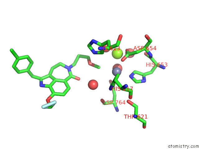
Mono view
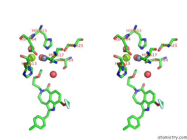
Stereo pair view
Mono view
Stereo pair view
A full contact list of Zinc with other atoms in the Zn binding
site number 1 of Crystal Structure of PDE5A in Complex with Inhibitor L12 within 5.0Å range:
|
Reference:
D.Wu,
X.Zheng,
R.Liu,
Z.Li,
Z.Jiang,
Q.Zhou,
Y.Huang,
X.N.Wu,
C.Zhang,
Y.Y.Huang,
H.B.Luo.
Free Energy Perturbation (Fep)-Guided Scaffold Hopping. Acta Pharm Sin B V. 12 1351 2022.
ISSN: ISSN 2211-3835
PubMed: 35530128
DOI: 10.1016/J.APSB.2021.09.027
Page generated: Tue Oct 29 20:17:12 2024
ISSN: ISSN 2211-3835
PubMed: 35530128
DOI: 10.1016/J.APSB.2021.09.027
Last articles
Zn in 9J0NZn in 9J0O
Zn in 9J0P
Zn in 9FJX
Zn in 9EKB
Zn in 9C0F
Zn in 9CAH
Zn in 9CH0
Zn in 9CH3
Zn in 9CH1