Zinc »
PDB 7doy-7dws »
7duf »
Zinc in PDB 7duf: Crystal Structure of VIM1 Phd Finger.
Enzymatic activity of Crystal Structure of VIM1 Phd Finger.
All present enzymatic activity of Crystal Structure of VIM1 Phd Finger.:
2.3.2.27;
2.3.2.27;
Protein crystallography data
The structure of Crystal Structure of VIM1 Phd Finger., PDB code: 7duf
was solved by
S.Abhishek,
W.Deeksha,
D.J.Patel,
E.Rajakumara,
with X-Ray Crystallography technique. A brief refinement statistics is given in the table below:
| Resolution Low / High (Å) | 32.30 / 2.61 |
| Space group | P 31 2 1 |
| Cell size a, b, c (Å), α, β, γ (°) | 74.59, 74.59, 58.88, 90, 90, 120 |
| R / Rfree (%) | 21.5 / 23.2 |
Zinc Binding Sites:
The binding sites of Zinc atom in the Crystal Structure of VIM1 Phd Finger.
(pdb code 7duf). This binding sites where shown within
5.0 Angstroms radius around Zinc atom.
In total 4 binding sites of Zinc where determined in the Crystal Structure of VIM1 Phd Finger., PDB code: 7duf:
Jump to Zinc binding site number: 1; 2; 3; 4;
In total 4 binding sites of Zinc where determined in the Crystal Structure of VIM1 Phd Finger., PDB code: 7duf:
Jump to Zinc binding site number: 1; 2; 3; 4;
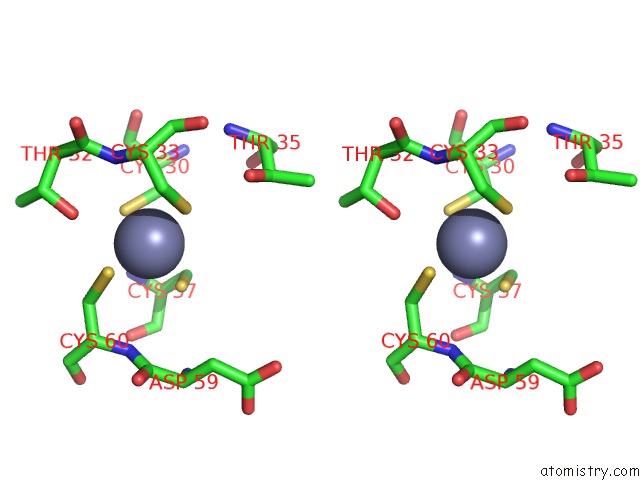
Zinc binding site 1 out of 4 in 7duf
Go back to
Zinc binding site 1 out
of 4 in the Crystal Structure of VIM1 Phd Finger.

Mono view

Stereo pair view
Mono view
Stereo pair view
A full contact list of Zinc with other atoms in the Zn binding
site number 1 of Crystal Structure of VIM1 Phd Finger. within 5.0Å range:
|
Zinc binding site 2 out of 4 in 7duf
Go back to
Zinc binding site 2 out
of 4 in the Crystal Structure of VIM1 Phd Finger.

Mono view

Stereo pair view
Mono view
Stereo pair view
A full contact list of Zinc with other atoms in the Zn binding
site number 2 of Crystal Structure of VIM1 Phd Finger. within 5.0Å range:
|
Zinc binding site 3 out of 4 in 7duf
Go back to
Zinc binding site 3 out
of 4 in the Crystal Structure of VIM1 Phd Finger.

Mono view
Stereo pair view
Mono view
Stereo pair view
A full contact list of Zinc with other atoms in the Zn binding
site number 3 of Crystal Structure of VIM1 Phd Finger. within 5.0Å range:
|
Zinc binding site 4 out of 4 in 7duf
Go back to
Zinc binding site 4 out
of 4 in the Crystal Structure of VIM1 Phd Finger.

Mono view

Stereo pair view
Mono view
Stereo pair view
A full contact list of Zinc with other atoms in the Zn binding
site number 4 of Crystal Structure of VIM1 Phd Finger. within 5.0Å range:
|
Reference:
S.Abhishek,
W.Deeksha,
E.Rajakumara.
Helical and Beta-Turn Conformations in the Peptide Recognition Regions of the VIM1 Phd Finger Abrogate H3K4 Peptide Recognition. Biochemistry 2021.
ISSN: ISSN 0006-2960
PubMed: 34404204
DOI: 10.1021/ACS.BIOCHEM.1C00191
Page generated: Tue Oct 29 19:19:34 2024
ISSN: ISSN 0006-2960
PubMed: 34404204
DOI: 10.1021/ACS.BIOCHEM.1C00191
Last articles
Zn in 9J0NZn in 9J0O
Zn in 9J0P
Zn in 9FJX
Zn in 9EKB
Zn in 9C0F
Zn in 9CAH
Zn in 9CH0
Zn in 9CH3
Zn in 9CH1