Zinc »
PDB 7del-7dox »
7diy »
Zinc in PDB 7diy: Crystal Structure of Sars-Cov-2 NSP10 Bound to NSP14-Exoribonuclease Domain
Protein crystallography data
The structure of Crystal Structure of Sars-Cov-2 NSP10 Bound to NSP14-Exoribonuclease Domain, PDB code: 7diy
was solved by
S.Lin,
H.Chen,
Z.M.Chen,
F.L.Yang,
F.Ye,
Y.Zheng,
J.Yang,
X.Lin,
H.L.Sun,
L.L.Wang,
A.Wen,
Y.Cao,
G.W.Lu,
with X-Ray Crystallography technique. A brief refinement statistics is given in the table below:
| Resolution Low / High (Å) | 42.77 / 2.69 |
| Space group | P 21 21 21 |
| Cell size a, b, c (Å), α, β, γ (°) | 53.929, 68.28, 109.739, 90, 90, 90 |
| R / Rfree (%) | 23 / 26.4 |
Other elements in 7diy:
The structure of Crystal Structure of Sars-Cov-2 NSP10 Bound to NSP14-Exoribonuclease Domain also contains other interesting chemical elements:
| Magnesium | (Mg) | 1 atom |
Zinc Binding Sites:
The binding sites of Zinc atom in the Crystal Structure of Sars-Cov-2 NSP10 Bound to NSP14-Exoribonuclease Domain
(pdb code 7diy). This binding sites where shown within
5.0 Angstroms radius around Zinc atom.
In total 4 binding sites of Zinc where determined in the Crystal Structure of Sars-Cov-2 NSP10 Bound to NSP14-Exoribonuclease Domain, PDB code: 7diy:
Jump to Zinc binding site number: 1; 2; 3; 4;
In total 4 binding sites of Zinc where determined in the Crystal Structure of Sars-Cov-2 NSP10 Bound to NSP14-Exoribonuclease Domain, PDB code: 7diy:
Jump to Zinc binding site number: 1; 2; 3; 4;
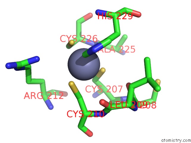
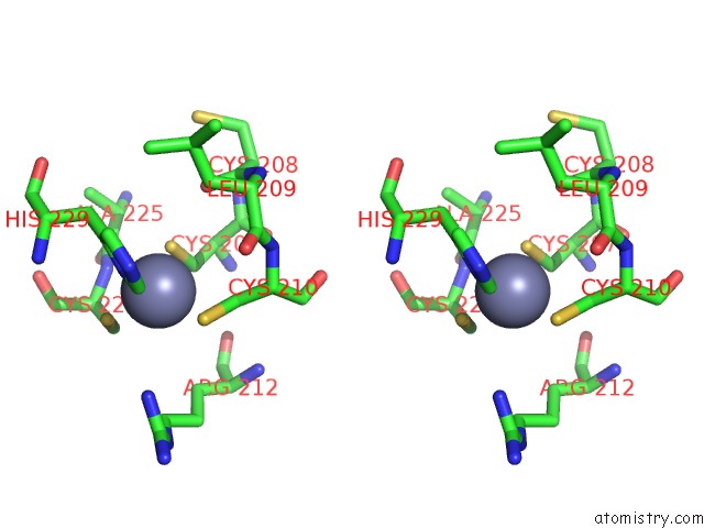
Zinc binding site 1 out of 4 in 7diy
Go back to
Zinc binding site 1 out
of 4 in the Crystal Structure of Sars-Cov-2 NSP10 Bound to NSP14-Exoribonuclease Domain
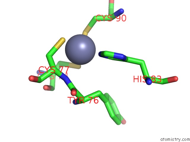
Mono view
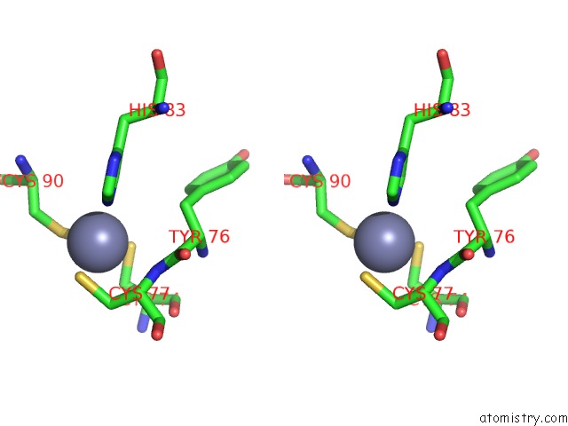
Stereo pair view
Mono view
Stereo pair view
A full contact list of Zinc with other atoms in the Zn binding
site number 1 of Crystal Structure of Sars-Cov-2 NSP10 Bound to NSP14-Exoribonuclease Domain within 5.0Å range:
|
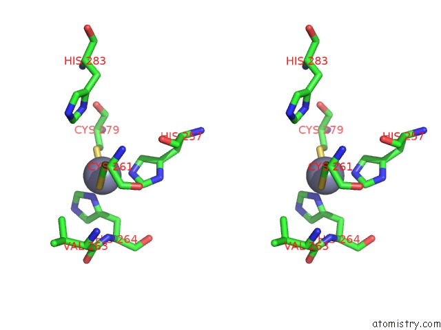
Zinc binding site 2 out of 4 in 7diy
Go back to
Zinc binding site 2 out
of 4 in the Crystal Structure of Sars-Cov-2 NSP10 Bound to NSP14-Exoribonuclease Domain
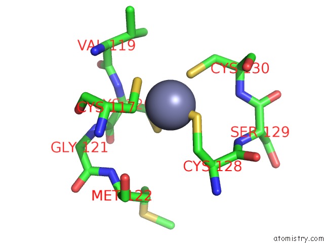
Mono view
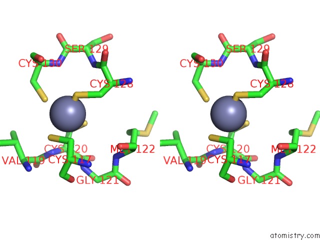
Stereo pair view
Mono view
Stereo pair view
A full contact list of Zinc with other atoms in the Zn binding
site number 2 of Crystal Structure of Sars-Cov-2 NSP10 Bound to NSP14-Exoribonuclease Domain within 5.0Å range:
|
Zinc binding site 3 out of 4 in 7diy
Go back to
Zinc binding site 3 out
of 4 in the Crystal Structure of Sars-Cov-2 NSP10 Bound to NSP14-Exoribonuclease Domain
Mono view
Stereo pair view
Mono view
Stereo pair view
A full contact list of Zinc with other atoms in the Zn binding
site number 3 of Crystal Structure of Sars-Cov-2 NSP10 Bound to NSP14-Exoribonuclease Domain within 5.0Å range:
|
Zinc binding site 4 out of 4 in 7diy
Go back to
Zinc binding site 4 out
of 4 in the Crystal Structure of Sars-Cov-2 NSP10 Bound to NSP14-Exoribonuclease Domain

Mono view
Stereo pair view
Mono view
Stereo pair view
A full contact list of Zinc with other atoms in the Zn binding
site number 4 of Crystal Structure of Sars-Cov-2 NSP10 Bound to NSP14-Exoribonuclease Domain within 5.0Å range:
|
Reference:
S.Lin,
H.Chen,
Z.Chen,
F.Yang,
F.Ye,
Y.Zheng,
J.Yang,
X.Lin,
H.Sun,
L.Wang,
A.Wen,
H.Dong,
Q.Xiao,
D.Deng,
Y.Cao,
G.Lu.
Crystal Structure of Sars-Cov-2 NSP10 Bound to NSP14-Exon Domain Reveals An Exoribonuclease with Both Structural and Functional Integrity. Nucleic Acids Res. 2021.
ISSN: ESSN 1362-4962
PubMed: 33956156
DOI: 10.1093/NAR/GKAB320
Page generated: Tue Oct 29 19:03:19 2024
ISSN: ESSN 1362-4962
PubMed: 33956156
DOI: 10.1093/NAR/GKAB320
Last articles
Zn in 9J0NZn in 9J0O
Zn in 9J0P
Zn in 9FJX
Zn in 9EKB
Zn in 9C0F
Zn in 9CAH
Zn in 9CH0
Zn in 9CH3
Zn in 9CH1