Zinc »
PDB 7del-7dox »
7dg2 »
Zinc in PDB 7dg2: NSE1-NSE3-NSE4 Complex
Enzymatic activity of NSE1-NSE3-NSE4 Complex
All present enzymatic activity of NSE1-NSE3-NSE4 Complex:
2.3.2.27;
2.3.2.27;
Protein crystallography data
The structure of NSE1-NSE3-NSE4 Complex, PDB code: 7dg2
was solved by
Y.Cho,
A.Jo,
with X-Ray Crystallography technique. A brief refinement statistics is given in the table below:
| Resolution Low / High (Å) | 41.59 / 1.70 |
| Space group | P 21 21 21 |
| Cell size a, b, c (Å), α, β, γ (°) | 53.486, 66.161, 168.367, 90, 90, 90 |
| R / Rfree (%) | 19 / 21.8 |
Zinc Binding Sites:
The binding sites of Zinc atom in the NSE1-NSE3-NSE4 Complex
(pdb code 7dg2). This binding sites where shown within
5.0 Angstroms radius around Zinc atom.
In total 2 binding sites of Zinc where determined in the NSE1-NSE3-NSE4 Complex, PDB code: 7dg2:
Jump to Zinc binding site number: 1; 2;
In total 2 binding sites of Zinc where determined in the NSE1-NSE3-NSE4 Complex, PDB code: 7dg2:
Jump to Zinc binding site number: 1; 2;
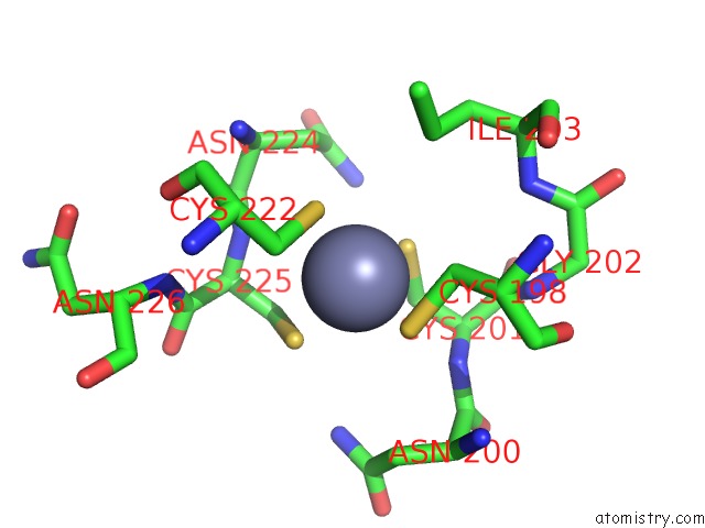
Zinc binding site 1 out of 2 in 7dg2
Go back to
Zinc binding site 1 out
of 2 in the NSE1-NSE3-NSE4 Complex

Mono view

Stereo pair view
Mono view
Stereo pair view
A full contact list of Zinc with other atoms in the Zn binding
site number 1 of NSE1-NSE3-NSE4 Complex within 5.0Å range:
|
Zinc binding site 2 out of 2 in 7dg2
Go back to
Zinc binding site 2 out
of 2 in the NSE1-NSE3-NSE4 Complex
Mono view

Stereo pair view
Mono view
Stereo pair view
A full contact list of Zinc with other atoms in the Zn binding
site number 2 of NSE1-NSE3-NSE4 Complex within 5.0Å range:
|
Reference:
A.Jo,
S.Li,
J.W.Shin,
X.Zhao,
Y.Cho.
Structure Basis For Shaping the NSE4 Protein By the NSE1 and NSE3 Dimer Within the SMC5/6 Complex. J.Mol.Biol. V. 433 66910 2021.
ISSN: ESSN 1089-8638
PubMed: 33676928
DOI: 10.1016/J.JMB.2021.166910
Page generated: Tue Oct 29 18:55:32 2024
ISSN: ESSN 1089-8638
PubMed: 33676928
DOI: 10.1016/J.JMB.2021.166910
Last articles
Zn in 9J0NZn in 9J0O
Zn in 9J0P
Zn in 9FJX
Zn in 9EKB
Zn in 9C0F
Zn in 9CAH
Zn in 9CH0
Zn in 9CH3
Zn in 9CH1