Zinc »
PDB 7c3s-7ci4 »
7c7l »
Zinc in PDB 7c7l: Cryo-Em Structure of the CAS12F1-Sgrna-Target Dna Complex
Zinc Binding Sites:
The binding sites of Zinc atom in the Cryo-Em Structure of the CAS12F1-Sgrna-Target Dna Complex
(pdb code 7c7l). This binding sites where shown within
5.0 Angstroms radius around Zinc atom.
In total 3 binding sites of Zinc where determined in the Cryo-Em Structure of the CAS12F1-Sgrna-Target Dna Complex, PDB code: 7c7l:
Jump to Zinc binding site number: 1; 2; 3;
In total 3 binding sites of Zinc where determined in the Cryo-Em Structure of the CAS12F1-Sgrna-Target Dna Complex, PDB code: 7c7l:
Jump to Zinc binding site number: 1; 2; 3;
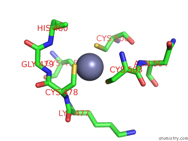
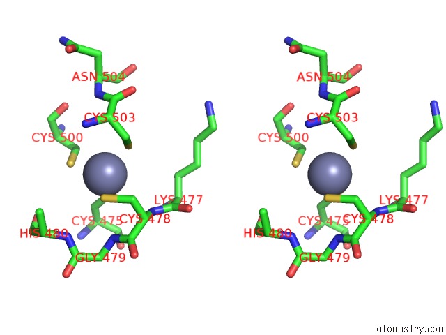
Zinc binding site 1 out of 3 in 7c7l
Go back to
Zinc binding site 1 out
of 3 in the Cryo-Em Structure of the CAS12F1-Sgrna-Target Dna Complex
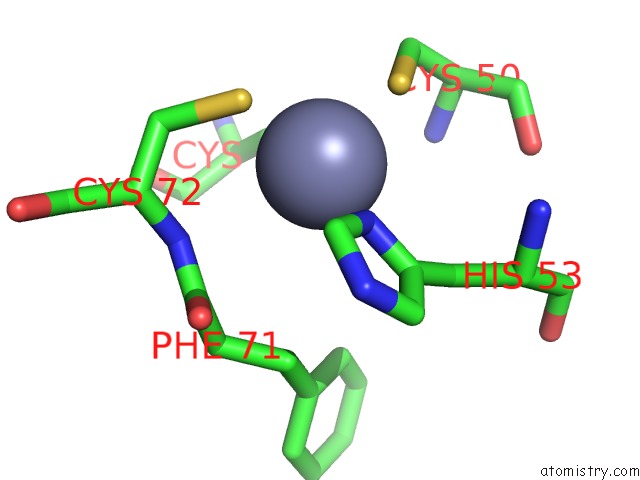
Mono view
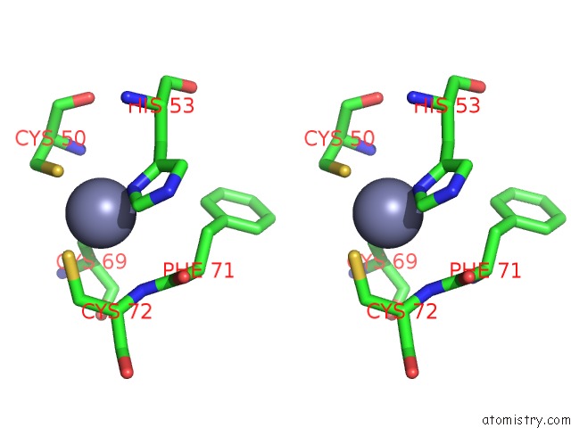
Stereo pair view
Mono view
Stereo pair view
A full contact list of Zinc with other atoms in the Zn binding
site number 1 of Cryo-Em Structure of the CAS12F1-Sgrna-Target Dna Complex within 5.0Å range:
|
Zinc binding site 2 out of 3 in 7c7l
Go back to
Zinc binding site 2 out
of 3 in the Cryo-Em Structure of the CAS12F1-Sgrna-Target Dna Complex
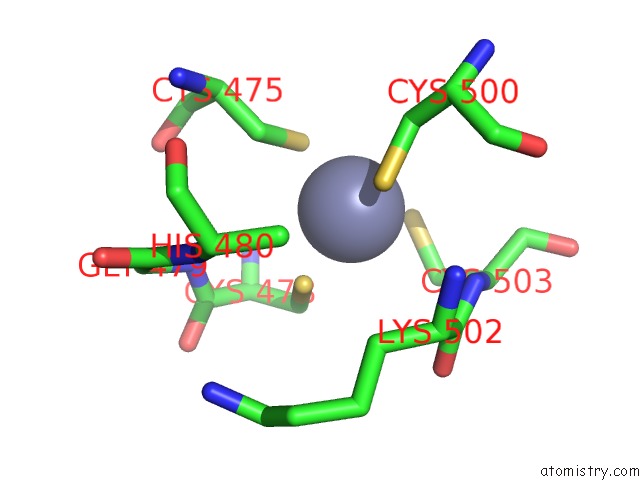
Mono view
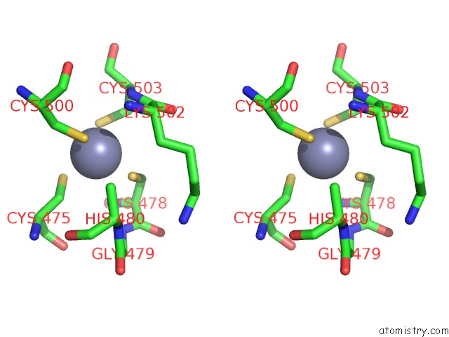
Stereo pair view
Mono view
Stereo pair view
A full contact list of Zinc with other atoms in the Zn binding
site number 2 of Cryo-Em Structure of the CAS12F1-Sgrna-Target Dna Complex within 5.0Å range:
|
Zinc binding site 3 out of 3 in 7c7l
Go back to
Zinc binding site 3 out
of 3 in the Cryo-Em Structure of the CAS12F1-Sgrna-Target Dna Complex
Mono view
Stereo pair view
Mono view
Stereo pair view
A full contact list of Zinc with other atoms in the Zn binding
site number 3 of Cryo-Em Structure of the CAS12F1-Sgrna-Target Dna Complex within 5.0Å range:
|
Reference:
S.N.Takeda,
R.Nakagawa,
S.Okazaki,
H.Hirano,
K.Kobayashi,
T.Kusakizako,
T.Nishizawa,
K.Yamashita,
H.Nishimasu,
O.Nureki.
Structure of the Miniature Type V-F Crispr-Cas Effector Enzyme Mol.Cell V. 81 2020.
ISSN: ISSN 1097-2765
DOI: 10.1016/J.MOLCEL.2020.11.035
Page generated: Tue Oct 29 18:04:56 2024
ISSN: ISSN 1097-2765
DOI: 10.1016/J.MOLCEL.2020.11.035
Last articles
Zn in 9J0NZn in 9J0O
Zn in 9J0P
Zn in 9FJX
Zn in 9EKB
Zn in 9C0F
Zn in 9CAH
Zn in 9CH0
Zn in 9CH3
Zn in 9CH1