Zinc »
PDB 7aez-7aoe »
7agl »
Zinc in PDB 7agl: Crystal Structure of the Apo Form of the N-Acetylmuramyl-L-Alanine Amidase, AMI1, From Mycobacterium Abscessus.
Protein crystallography data
The structure of Crystal Structure of the Apo Form of the N-Acetylmuramyl-L-Alanine Amidase, AMI1, From Mycobacterium Abscessus., PDB code: 7agl
was solved by
M.Blaise,
with X-Ray Crystallography technique. A brief refinement statistics is given in the table below:
| Resolution Low / High (Å) | 38.42 / 1.60 |
| Space group | P 41 21 2 |
| Cell size a, b, c (Å), α, β, γ (°) | 85.920, 85.920, 73.660, 90.00, 90.00, 90.00 |
| R / Rfree (%) | 16.8 / 19.6 |
Zinc Binding Sites:
The binding sites of Zinc atom in the Crystal Structure of the Apo Form of the N-Acetylmuramyl-L-Alanine Amidase, AMI1, From Mycobacterium Abscessus.
(pdb code 7agl). This binding sites where shown within
5.0 Angstroms radius around Zinc atom.
In total only one binding site of Zinc was determined in the Crystal Structure of the Apo Form of the N-Acetylmuramyl-L-Alanine Amidase, AMI1, From Mycobacterium Abscessus., PDB code: 7agl:
In total only one binding site of Zinc was determined in the Crystal Structure of the Apo Form of the N-Acetylmuramyl-L-Alanine Amidase, AMI1, From Mycobacterium Abscessus., PDB code: 7agl:
Zinc binding site 1 out of 1 in 7agl
Go back to
Zinc binding site 1 out
of 1 in the Crystal Structure of the Apo Form of the N-Acetylmuramyl-L-Alanine Amidase, AMI1, From Mycobacterium Abscessus.
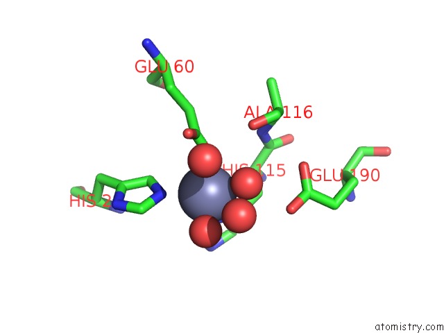
Mono view
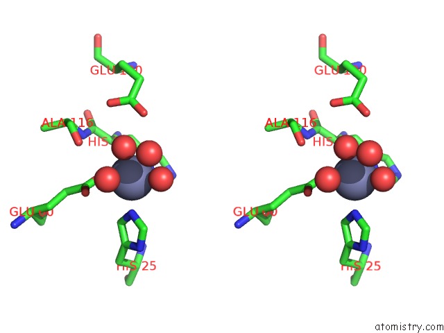
Stereo pair view
Mono view
Stereo pair view
A full contact list of Zinc with other atoms in the Zn binding
site number 1 of Crystal Structure of the Apo Form of the N-Acetylmuramyl-L-Alanine Amidase, AMI1, From Mycobacterium Abscessus. within 5.0Å range:
|
Reference:
T.Kussau,
N.Van Wyk,
M.D.Johansen,
H.M.A.B.Alsarraf,
A.Neyret,
C.Hamela,
K.K.Sorensen,
M.B.Thygesen,
C.Beauvineau,
L.Kremer,
M.Blaise.
Functional Characterization of the N -Acetylmuramyl-L-Alanine Amidase, AMI1, From Mycobacterium Abscessus . Cells V. 9 2020.
ISSN: ESSN 2073-4409
PubMed: 33158165
DOI: 10.3390/CELLS9112410
Page generated: Tue Oct 29 16:49:12 2024
ISSN: ESSN 2073-4409
PubMed: 33158165
DOI: 10.3390/CELLS9112410
Last articles
Zn in 9J0NZn in 9J0O
Zn in 9J0P
Zn in 9FJX
Zn in 9EKB
Zn in 9C0F
Zn in 9CAH
Zn in 9CH0
Zn in 9CH3
Zn in 9CH1