Zinc »
PDB 6zki-6zpu »
6zpc »
Zinc in PDB 6zpc: Cyanophage S-2L Hd Phosphohydrolase (Datz) Bound to Datp
Protein crystallography data
The structure of Cyanophage S-2L Hd Phosphohydrolase (Datz) Bound to Datp, PDB code: 6zpc
was solved by
D.Czernecki,
P.Legrand,
M.Delarue,
with X-Ray Crystallography technique. A brief refinement statistics is given in the table below:
| Resolution Low / High (Å) | 40.42 / 1.27 |
| Space group | H 3 2 |
| Cell size a, b, c (Å), α, β, γ (°) | 141.65, 141.65, 53.73, 90, 90, 120 |
| R / Rfree (%) | 12.6 / 14.6 |
Zinc Binding Sites:
The binding sites of Zinc atom in the Cyanophage S-2L Hd Phosphohydrolase (Datz) Bound to Datp
(pdb code 6zpc). This binding sites where shown within
5.0 Angstroms radius around Zinc atom.
In total only one binding site of Zinc was determined in the Cyanophage S-2L Hd Phosphohydrolase (Datz) Bound to Datp, PDB code: 6zpc:
In total only one binding site of Zinc was determined in the Cyanophage S-2L Hd Phosphohydrolase (Datz) Bound to Datp, PDB code: 6zpc:
Zinc binding site 1 out of 1 in 6zpc
Go back to
Zinc binding site 1 out
of 1 in the Cyanophage S-2L Hd Phosphohydrolase (Datz) Bound to Datp
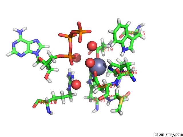
Mono view
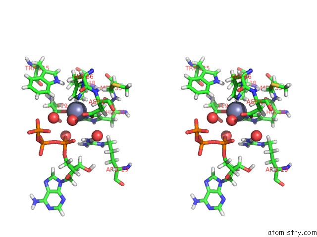
Stereo pair view
Mono view
Stereo pair view
A full contact list of Zinc with other atoms in the Zn binding
site number 1 of Cyanophage S-2L Hd Phosphohydrolase (Datz) Bound to Datp within 5.0Å range:
|
Reference:
D.Czernecki,
P.Legrand,
M.Tekpinar,
S.Rosario,
P.A.Kaminski,
M.Delarue.
How Cyanophage S-2L Rejects Adenine and Incorporates 2-Aminoadenine to Saturate Hydrogen Bonding in Its Dna Nat Commun 2021.
ISSN: ESSN 2041-1723
Page generated: Tue Oct 29 15:51:28 2024
ISSN: ESSN 2041-1723
Last articles
Zn in 9J0NZn in 9J0O
Zn in 9J0P
Zn in 9FJX
Zn in 9EKB
Zn in 9C0F
Zn in 9CAH
Zn in 9CH0
Zn in 9CH3
Zn in 9CH1