Zinc »
PDB 6zki-6zpu »
6znc »
Zinc in PDB 6znc: Structural Basis of Reactivation of Oncogenic P53 Mutants By A Small Molecule: Methylene Quinuclidinone (Mq). Human Wild-Type P53DBD Bound to Dna and Mq: Wt-Dna-Mq (I)
Protein crystallography data
The structure of Structural Basis of Reactivation of Oncogenic P53 Mutants By A Small Molecule: Methylene Quinuclidinone (Mq). Human Wild-Type P53DBD Bound to Dna and Mq: Wt-Dna-Mq (I), PDB code: 6znc
was solved by
H.Rozenberg,
O.Degtjarik,
Y.Diskin-Posner,
Z.Shakked,
with X-Ray Crystallography technique. A brief refinement statistics is given in the table below:
| Resolution Low / High (Å) | 46.61 / 1.64 |
| Space group | C 1 2 1 |
| Cell size a, b, c (Å), α, β, γ (°) | 137.744, 49.537, 34.066, 90, 92.79, 90 |
| R / Rfree (%) | 16.1 / 19 |
Zinc Binding Sites:
The binding sites of Zinc atom in the Structural Basis of Reactivation of Oncogenic P53 Mutants By A Small Molecule: Methylene Quinuclidinone (Mq). Human Wild-Type P53DBD Bound to Dna and Mq: Wt-Dna-Mq (I)
(pdb code 6znc). This binding sites where shown within
5.0 Angstroms radius around Zinc atom.
In total only one binding site of Zinc was determined in the Structural Basis of Reactivation of Oncogenic P53 Mutants By A Small Molecule: Methylene Quinuclidinone (Mq). Human Wild-Type P53DBD Bound to Dna and Mq: Wt-Dna-Mq (I), PDB code: 6znc:
In total only one binding site of Zinc was determined in the Structural Basis of Reactivation of Oncogenic P53 Mutants By A Small Molecule: Methylene Quinuclidinone (Mq). Human Wild-Type P53DBD Bound to Dna and Mq: Wt-Dna-Mq (I), PDB code: 6znc:
Zinc binding site 1 out of 1 in 6znc
Go back to
Zinc binding site 1 out
of 1 in the Structural Basis of Reactivation of Oncogenic P53 Mutants By A Small Molecule: Methylene Quinuclidinone (Mq). Human Wild-Type P53DBD Bound to Dna and Mq: Wt-Dna-Mq (I)
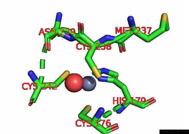
Mono view
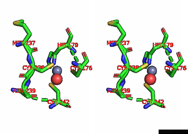
Stereo pair view
Mono view
Stereo pair view
A full contact list of Zinc with other atoms in the Zn binding
site number 1 of Structural Basis of Reactivation of Oncogenic P53 Mutants By A Small Molecule: Methylene Quinuclidinone (Mq). Human Wild-Type P53DBD Bound to Dna and Mq: Wt-Dna-Mq (I) within 5.0Å range:
|
Reference:
O.Degtjarik,
D.Golovenko,
Y.Diskin-Posner,
L.Abrahmsen,
H.Rozenberg,
Z.Shakked.
P53 Structure 11 To Be Published.
Page generated: Tue Oct 29 15:47:09 2024
Last articles
Zn in 9J0NZn in 9J0O
Zn in 9J0P
Zn in 9FJX
Zn in 9EKB
Zn in 9C0F
Zn in 9CAH
Zn in 9CH0
Zn in 9CH3
Zn in 9CH1