Zinc »
PDB 6wdv-6wq1 »
6wdy »
Zinc in PDB 6wdy: Crystal Structure of Danio Rerio Histone Deacetylase 10 in Complex with Indole Phenylhydroxamate Inhibitor
Enzymatic activity of Crystal Structure of Danio Rerio Histone Deacetylase 10 in Complex with Indole Phenylhydroxamate Inhibitor
All present enzymatic activity of Crystal Structure of Danio Rerio Histone Deacetylase 10 in Complex with Indole Phenylhydroxamate Inhibitor:
3.5.1.48; 3.5.1.62;
3.5.1.48; 3.5.1.62;
Protein crystallography data
The structure of Crystal Structure of Danio Rerio Histone Deacetylase 10 in Complex with Indole Phenylhydroxamate Inhibitor, PDB code: 6wdy
was solved by
C.J.Herbst-Gervasoni,
D.W.Christianson,
with X-Ray Crystallography technique. A brief refinement statistics is given in the table below:
| Resolution Low / High (Å) | 69.86 / 2.65 |
| Space group | P 31 2 1 |
| Cell size a, b, c (Å), α, β, γ (°) | 80.670, 80.670, 243.980, 90.00, 90.00, 120.00 |
| R / Rfree (%) | 19 / 25.5 |
Other elements in 6wdy:
The structure of Crystal Structure of Danio Rerio Histone Deacetylase 10 in Complex with Indole Phenylhydroxamate Inhibitor also contains other interesting chemical elements:
| Potassium | (K) | 2 atoms |
Zinc Binding Sites:
The binding sites of Zinc atom in the Crystal Structure of Danio Rerio Histone Deacetylase 10 in Complex with Indole Phenylhydroxamate Inhibitor
(pdb code 6wdy). This binding sites where shown within
5.0 Angstroms radius around Zinc atom.
In total only one binding site of Zinc was determined in the Crystal Structure of Danio Rerio Histone Deacetylase 10 in Complex with Indole Phenylhydroxamate Inhibitor, PDB code: 6wdy:
In total only one binding site of Zinc was determined in the Crystal Structure of Danio Rerio Histone Deacetylase 10 in Complex with Indole Phenylhydroxamate Inhibitor, PDB code: 6wdy:
Zinc binding site 1 out of 1 in 6wdy
Go back to
Zinc binding site 1 out
of 1 in the Crystal Structure of Danio Rerio Histone Deacetylase 10 in Complex with Indole Phenylhydroxamate Inhibitor
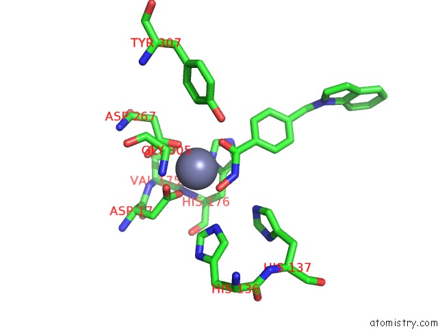
Mono view
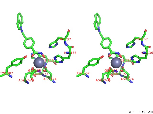
Stereo pair view
Mono view
Stereo pair view
A full contact list of Zinc with other atoms in the Zn binding
site number 1 of Crystal Structure of Danio Rerio Histone Deacetylase 10 in Complex with Indole Phenylhydroxamate Inhibitor within 5.0Å range:
|
Reference:
C.J.Herbst-Gervasoni,
R.R.Steimbach,
M.Morgen,
A.K.Miller,
D.W.Christianson.
Structural Basis For the Selective Inhibition of HDAC10, the Cytosolic Polyamine Deacetylase. Acs Chem.Biol. V. 15 2154 2020.
ISSN: ESSN 1554-8937
PubMed: 32659072
DOI: 10.1021/ACSCHEMBIO.0C00362
Page generated: Tue Oct 29 09:18:52 2024
ISSN: ESSN 1554-8937
PubMed: 32659072
DOI: 10.1021/ACSCHEMBIO.0C00362
Last articles
Zn in 9J0NZn in 9J0O
Zn in 9J0P
Zn in 9FJX
Zn in 9EKB
Zn in 9C0F
Zn in 9CAH
Zn in 9CH0
Zn in 9CH3
Zn in 9CH1