Zinc »
PDB 6v77-6vpc »
6vhf »
Zinc in PDB 6vhf: Crystal Structure of RBBP5 Interacting Domain of CFP1
Protein crystallography data
The structure of Crystal Structure of RBBP5 Interacting Domain of CFP1, PDB code: 6vhf
was solved by
M.Joshi,
J.F.Couture,
with X-Ray Crystallography technique. A brief refinement statistics is given in the table below:
| Resolution Low / High (Å) | 49.03 / 2.31 |
| Space group | I 2 2 2 |
| Cell size a, b, c (Å), α, β, γ (°) | 67.266, 71.614, 78.132, 90.00, 90.00, 90.00 |
| R / Rfree (%) | 23.3 / 27.3 |
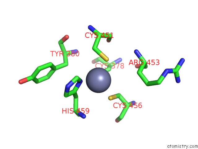
Zinc Binding Sites:
The binding sites of Zinc atom in the Crystal Structure of RBBP5 Interacting Domain of CFP1
(pdb code 6vhf). This binding sites where shown within
5.0 Angstroms radius around Zinc atom.
In total only one binding site of Zinc was determined in the Crystal Structure of RBBP5 Interacting Domain of CFP1, PDB code: 6vhf:
In total only one binding site of Zinc was determined in the Crystal Structure of RBBP5 Interacting Domain of CFP1, PDB code: 6vhf:
Zinc binding site 1 out of 1 in 6vhf
Go back to
Zinc binding site 1 out
of 1 in the Crystal Structure of RBBP5 Interacting Domain of CFP1
Mono view

Stereo pair view
Mono view
Stereo pair view
A full contact list of Zinc with other atoms in the Zn binding
site number 1 of Crystal Structure of RBBP5 Interacting Domain of CFP1 within 5.0Å range:
|
Reference:
Y.Yang,
M.Joshi,
Y.H.Takahashi,
Z.Ning,
Q.Qu,
J.S.Brunzelle,
G.Skiniotis,
D.Figeys,
A.Shilatifard,
J.F.Couture.
A Non-Canonical Monovalent Zinc Finger Stabilizes the Integration of CFP1 Into the H3K4 Methyltransferase Complex Compass. Nucleic Acids Res. V. 48 421 2020.
ISSN: ESSN 1362-4962
PubMed: 31724694
DOI: 10.1093/NAR/GKZ1037
Page generated: Tue Oct 29 09:03:19 2024
ISSN: ESSN 1362-4962
PubMed: 31724694
DOI: 10.1093/NAR/GKZ1037
Last articles
Zn in 9J0NZn in 9J0O
Zn in 9J0P
Zn in 9FJX
Zn in 9EKB
Zn in 9C0F
Zn in 9CAH
Zn in 9CH0
Zn in 9CH3
Zn in 9CH1