Zinc in PDB 6uat: Crystal Structure of A GH128 (Subgroup I) Endo-Beta-1,3-Glucanase (E102A Mutant) From Amycolatopsis Mediterranei (AMGH128_I) in Complex with Laminaripentaose
Protein crystallography data
The structure of Crystal Structure of A GH128 (Subgroup I) Endo-Beta-1,3-Glucanase (E102A Mutant) From Amycolatopsis Mediterranei (AMGH128_I) in Complex with Laminaripentaose, PDB code: 6uat
was solved by
P.S.Vieira,
L.Cabral,
P.A.C.R.Costa,
C.R.Santos,
M.T.Murakami,
with X-Ray Crystallography technique. A brief refinement statistics is given in the table below:
| Resolution Low / High (Å) | 39.63 / 1.90 |
| Space group | P 1 21 1 |
| Cell size a, b, c (Å), α, β, γ (°) | 38.458, 79.138, 46.779, 90.00, 102.13, 90.00 |
| R / Rfree (%) | 18.5 / 23.6 |
Zinc Binding Sites:
The binding sites of Zinc atom in the Crystal Structure of A GH128 (Subgroup I) Endo-Beta-1,3-Glucanase (E102A Mutant) From Amycolatopsis Mediterranei (AMGH128_I) in Complex with Laminaripentaose
(pdb code 6uat). This binding sites where shown within
5.0 Angstroms radius around Zinc atom.
In total only one binding site of Zinc was determined in the Crystal Structure of A GH128 (Subgroup I) Endo-Beta-1,3-Glucanase (E102A Mutant) From Amycolatopsis Mediterranei (AMGH128_I) in Complex with Laminaripentaose, PDB code: 6uat:
In total only one binding site of Zinc was determined in the Crystal Structure of A GH128 (Subgroup I) Endo-Beta-1,3-Glucanase (E102A Mutant) From Amycolatopsis Mediterranei (AMGH128_I) in Complex with Laminaripentaose, PDB code: 6uat:
Zinc binding site 1 out of 1 in 6uat
Go back to
Zinc binding site 1 out
of 1 in the Crystal Structure of A GH128 (Subgroup I) Endo-Beta-1,3-Glucanase (E102A Mutant) From Amycolatopsis Mediterranei (AMGH128_I) in Complex with Laminaripentaose
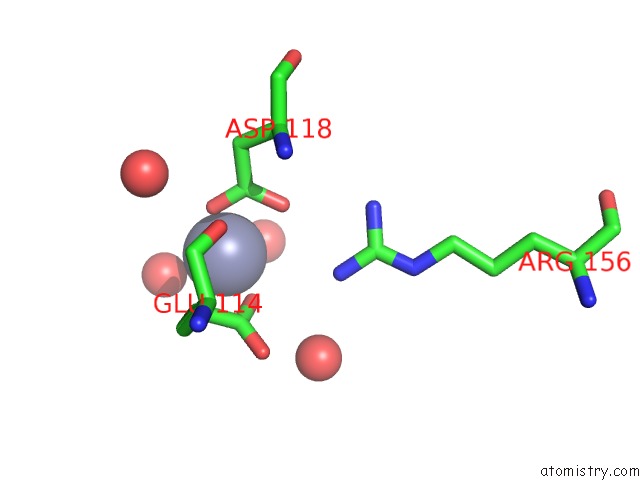
Mono view
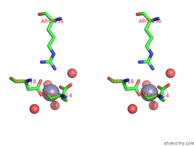
Stereo pair view
Mono view
Stereo pair view
A full contact list of Zinc with other atoms in the Zn binding
site number 1 of Crystal Structure of A GH128 (Subgroup I) Endo-Beta-1,3-Glucanase (E102A Mutant) From Amycolatopsis Mediterranei (AMGH128_I) in Complex with Laminaripentaose within 5.0Å range:
|
Reference:
C.R.Santos,
E.A.Lima,
F.Mandelli,
M.T.Murakami.
Structural Insights Into Beta-1,3-Glucan Cleavage By A Glycoside Hydrolase Family Nat.Chem.Biol. 2020.
ISSN: ESSN 1552-4469
DOI: 10.1038/S41589-020-0554-5
Page generated: Tue Oct 29 08:32:33 2024
ISSN: ESSN 1552-4469
DOI: 10.1038/S41589-020-0554-5
Last articles
Zn in 9MJ5Zn in 9HNW
Zn in 9G0L
Zn in 9FNE
Zn in 9DZN
Zn in 9E0I
Zn in 9D32
Zn in 9DAK
Zn in 8ZXC
Zn in 8ZUF