Zinc »
PDB 6t6r-6tm5 »
6thh »
Zinc in PDB 6thh: Crystal Structure of Type I-D Crispr-Cas Nuclease CAS10D in Complex with the SIRV3 ACRID1 (GP02) Anti-Crispr Protein
Protein crystallography data
The structure of Crystal Structure of Type I-D Crispr-Cas Nuclease CAS10D in Complex with the SIRV3 ACRID1 (GP02) Anti-Crispr Protein, PDB code: 6thh
was solved by
M.C.Manav,
D.E.Brodersen,
with X-Ray Crystallography technique. A brief refinement statistics is given in the table below:
| Resolution Low / High (Å) | 67.54 / 3.48 |
| Space group | P 43 21 2 |
| Cell size a, b, c (Å), α, β, γ (°) | 157.990, 157.990, 130.240, 90.00, 90.00, 90.00 |
| R / Rfree (%) | 23.6 / 25.4 |
Zinc Binding Sites:
The binding sites of Zinc atom in the Crystal Structure of Type I-D Crispr-Cas Nuclease CAS10D in Complex with the SIRV3 ACRID1 (GP02) Anti-Crispr Protein
(pdb code 6thh). This binding sites where shown within
5.0 Angstroms radius around Zinc atom.
In total only one binding site of Zinc was determined in the Crystal Structure of Type I-D Crispr-Cas Nuclease CAS10D in Complex with the SIRV3 ACRID1 (GP02) Anti-Crispr Protein, PDB code: 6thh:
In total only one binding site of Zinc was determined in the Crystal Structure of Type I-D Crispr-Cas Nuclease CAS10D in Complex with the SIRV3 ACRID1 (GP02) Anti-Crispr Protein, PDB code: 6thh:
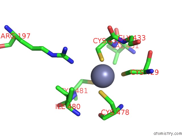
Zinc binding site 1 out of 1 in 6thh
Go back to
Zinc binding site 1 out
of 1 in the Crystal Structure of Type I-D Crispr-Cas Nuclease CAS10D in Complex with the SIRV3 ACRID1 (GP02) Anti-Crispr Protein
Mono view

Stereo pair view
Mono view
Stereo pair view
A full contact list of Zinc with other atoms in the Zn binding
site number 1 of Crystal Structure of Type I-D Crispr-Cas Nuclease CAS10D in Complex with the SIRV3 ACRID1 (GP02) Anti-Crispr Protein within 5.0Å range:
|
Reference:
M.C.Manav,
L.B.Van,
J.Lin,
A.Fuglsang,
X.Peng,
D.E.Brodersen.
Structural Basis For Inhibition of An Archaeal Crispr-Cas Type I-D Large Subunit By An Anti-Crispr Protein. Nat Commun V. 11 5993 2020.
ISSN: ESSN 2041-1723
PubMed: 33239638
DOI: 10.1038/S41467-020-19847-X
Page generated: Tue Oct 29 07:58:22 2024
ISSN: ESSN 2041-1723
PubMed: 33239638
DOI: 10.1038/S41467-020-19847-X
Last articles
Zn in 9MJ5Zn in 9HNW
Zn in 9G0L
Zn in 9FNE
Zn in 9DZN
Zn in 9E0I
Zn in 9D32
Zn in 9DAK
Zn in 8ZXC
Zn in 8ZUF