Zinc »
PDB 6sj2-6ssc »
6sno »
Zinc in PDB 6sno: Crystal Structures of Human PGM1 Isoform 2
Enzymatic activity of Crystal Structures of Human PGM1 Isoform 2
All present enzymatic activity of Crystal Structures of Human PGM1 Isoform 2:
5.4.2.2;
5.4.2.2;
Protein crystallography data
The structure of Crystal Structures of Human PGM1 Isoform 2, PDB code: 6sno
was solved by
P.H.Backe,
J.K.Laerdahl,
L.S.Kittelsen,
B.Dalhus,
L.Morkrid,
M.Bjoras,
with X-Ray Crystallography technique. A brief refinement statistics is given in the table below:
| Resolution Low / High (Å) | 43.57 / 2.70 |
| Space group | P 1 21 1 |
| Cell size a, b, c (Å), α, β, γ (°) | 72.954, 53.351, 76.321, 90.00, 98.44, 90.00 |
| R / Rfree (%) | 19.7 / 24.8 |
Zinc Binding Sites:
The binding sites of Zinc atom in the Crystal Structures of Human PGM1 Isoform 2
(pdb code 6sno). This binding sites where shown within
5.0 Angstroms radius around Zinc atom.
In total only one binding site of Zinc was determined in the Crystal Structures of Human PGM1 Isoform 2, PDB code: 6sno:
In total only one binding site of Zinc was determined in the Crystal Structures of Human PGM1 Isoform 2, PDB code: 6sno:
Zinc binding site 1 out of 1 in 6sno
Go back to
Zinc binding site 1 out
of 1 in the Crystal Structures of Human PGM1 Isoform 2
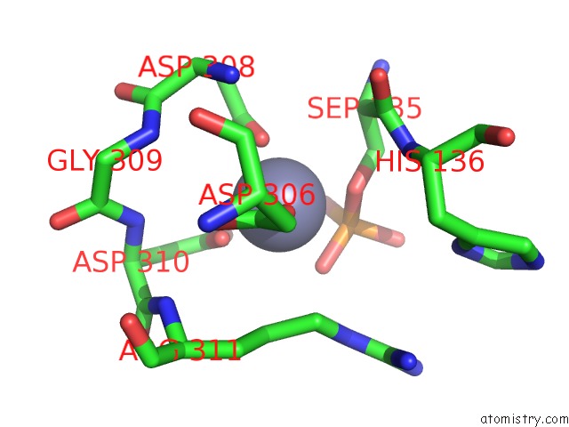
Mono view
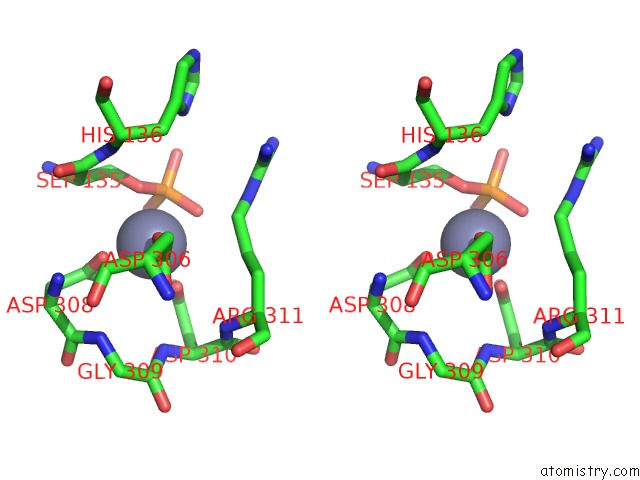
Stereo pair view
Mono view
Stereo pair view
A full contact list of Zinc with other atoms in the Zn binding
site number 1 of Crystal Structures of Human PGM1 Isoform 2 within 5.0Å range:
|
Reference:
P.H.Backe,
J.K.Laerdahl,
L.S.Kittelsen,
B.Dalhus,
L.Morkrid,
M.Bjoras.
Structural Basis For Substrate and Product Recognition in Human Phosphoglucomutase-1 (PGM1) Isoform 2, A Member of the Alpha-D-Phosphohexomutase Superfamily. Sci Rep V. 10 5656 2020.
ISSN: ESSN 2045-2322
PubMed: 32221390
DOI: 10.1038/S41598-020-62548-0
Page generated: Tue Oct 29 07:30:27 2024
ISSN: ESSN 2045-2322
PubMed: 32221390
DOI: 10.1038/S41598-020-62548-0
Last articles
Zn in 9J0NZn in 9J0O
Zn in 9J0P
Zn in 9FJX
Zn in 9EKB
Zn in 9C0F
Zn in 9CAH
Zn in 9CH0
Zn in 9CH3
Zn in 9CH1