Zinc »
PDB 6rwo-6s6b »
6ryo »
Zinc in PDB 6ryo: Bacterial Membrane Enzyme Structure By the in Meso Method at 1.9 A Resolution
Enzymatic activity of Bacterial Membrane Enzyme Structure By the in Meso Method at 1.9 A Resolution
All present enzymatic activity of Bacterial Membrane Enzyme Structure By the in Meso Method at 1.9 A Resolution:
3.4.23.36;
3.4.23.36;
Protein crystallography data
The structure of Bacterial Membrane Enzyme Structure By the in Meso Method at 1.9 A Resolution, PDB code: 6ryo
was solved by
C.Y.Huang,
S.Olatunji,
J.Bailey,
X.Yu,
V.Olieric,
M.Wang,
M.Caffrey,
with X-Ray Crystallography technique. A brief refinement statistics is given in the table below:
| Resolution Low / High (Å) | 45.26 / 1.92 |
| Space group | P 32 2 1 |
| Cell size a, b, c (Å), α, β, γ (°) | 52.258, 52.258, 135.882, 90.00, 90.00, 120.00 |
| R / Rfree (%) | 25 / 27.6 |
Zinc Binding Sites:
The binding sites of Zinc atom in the Bacterial Membrane Enzyme Structure By the in Meso Method at 1.9 A Resolution
(pdb code 6ryo). This binding sites where shown within
5.0 Angstroms radius around Zinc atom.
In total only one binding site of Zinc was determined in the Bacterial Membrane Enzyme Structure By the in Meso Method at 1.9 A Resolution, PDB code: 6ryo:
In total only one binding site of Zinc was determined in the Bacterial Membrane Enzyme Structure By the in Meso Method at 1.9 A Resolution, PDB code: 6ryo:
Zinc binding site 1 out of 1 in 6ryo
Go back to
Zinc binding site 1 out
of 1 in the Bacterial Membrane Enzyme Structure By the in Meso Method at 1.9 A Resolution
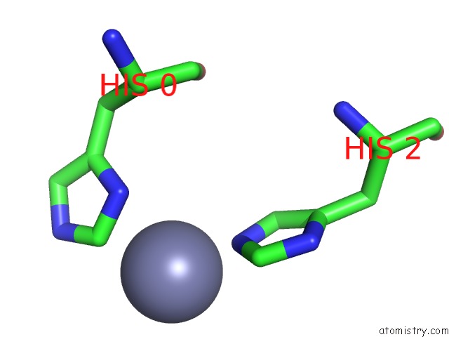
Mono view
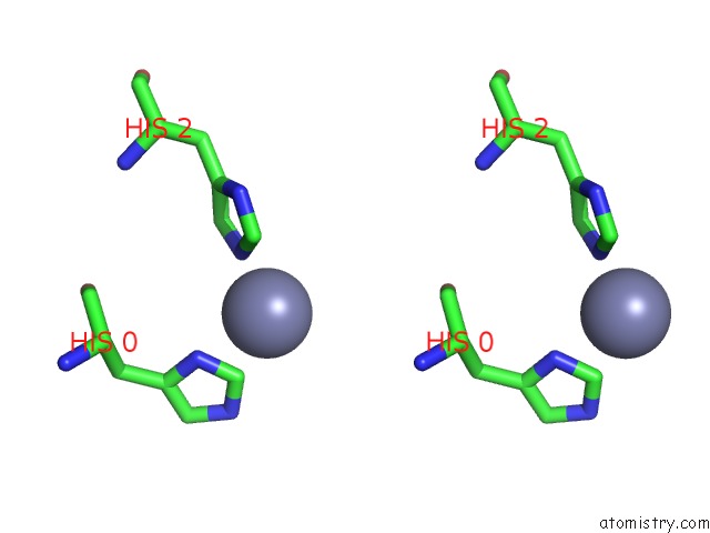
Stereo pair view
Mono view
Stereo pair view
A full contact list of Zinc with other atoms in the Zn binding
site number 1 of Bacterial Membrane Enzyme Structure By the in Meso Method at 1.9 A Resolution within 5.0Å range:
|
Reference:
C.Y.Huang,
S.Olatunji,
J.Bailey,
X.Yu,
V.Olieric,
M.Wang,
M.Caffrey.
Structures of Lipoprotein Signal Peptidase II From Staphylococcus Aureus Complexed with Antibiotics Globomycin and Myxovirescin Nat Commun.
ISSN: ESSN 2041-1723
DOI: 10.1038/S41467-019-13724-Y
Page generated: Tue Oct 29 06:55:53 2024
ISSN: ESSN 2041-1723
DOI: 10.1038/S41467-019-13724-Y
Last articles
Zn in 9J0NZn in 9J0O
Zn in 9J0P
Zn in 9FJX
Zn in 9EKB
Zn in 9C0F
Zn in 9CAH
Zn in 9CH0
Zn in 9CH3
Zn in 9CH1