Zinc »
PDB 6rpn-6rwn »
6rps »
Zinc in PDB 6rps: X-Ray Crystal Structure of Carbonic Anhydrase XII Complexed with A Theranostic Monoclonal Antibody Fragment
Enzymatic activity of X-Ray Crystal Structure of Carbonic Anhydrase XII Complexed with A Theranostic Monoclonal Antibody Fragment
All present enzymatic activity of X-Ray Crystal Structure of Carbonic Anhydrase XII Complexed with A Theranostic Monoclonal Antibody Fragment:
4.2.1.1;
4.2.1.1;
Protein crystallography data
The structure of X-Ray Crystal Structure of Carbonic Anhydrase XII Complexed with A Theranostic Monoclonal Antibody Fragment, PDB code: 6rps
was solved by
V.Alterio,
D.Esposito,
G.De Simone,
with X-Ray Crystallography technique. A brief refinement statistics is given in the table below:
| Resolution Low / High (Å) | 49.65 / 2.79 |
| Space group | I 2 2 2 |
| Cell size a, b, c (Å), α, β, γ (°) | 77.335, 222.165, 266.988, 90.00, 90.00, 90.00 |
| R / Rfree (%) | 20.3 / 23.1 |
Other elements in 6rps:
The structure of X-Ray Crystal Structure of Carbonic Anhydrase XII Complexed with A Theranostic Monoclonal Antibody Fragment also contains other interesting chemical elements:
| Cadmium | (Cd) | 4 atoms |
| Chlorine | (Cl) | 1 atom |
Zinc Binding Sites:
The binding sites of Zinc atom in the X-Ray Crystal Structure of Carbonic Anhydrase XII Complexed with A Theranostic Monoclonal Antibody Fragment
(pdb code 6rps). This binding sites where shown within
5.0 Angstroms radius around Zinc atom.
In total 2 binding sites of Zinc where determined in the X-Ray Crystal Structure of Carbonic Anhydrase XII Complexed with A Theranostic Monoclonal Antibody Fragment, PDB code: 6rps:
Jump to Zinc binding site number: 1; 2;
In total 2 binding sites of Zinc where determined in the X-Ray Crystal Structure of Carbonic Anhydrase XII Complexed with A Theranostic Monoclonal Antibody Fragment, PDB code: 6rps:
Jump to Zinc binding site number: 1; 2;
Zinc binding site 1 out of 2 in 6rps
Go back to
Zinc binding site 1 out
of 2 in the X-Ray Crystal Structure of Carbonic Anhydrase XII Complexed with A Theranostic Monoclonal Antibody Fragment
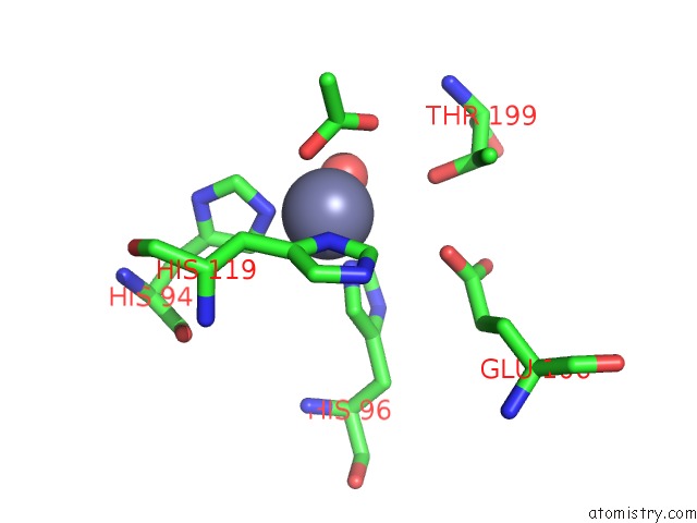
Mono view
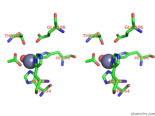
Stereo pair view
Mono view
Stereo pair view
A full contact list of Zinc with other atoms in the Zn binding
site number 1 of X-Ray Crystal Structure of Carbonic Anhydrase XII Complexed with A Theranostic Monoclonal Antibody Fragment within 5.0Å range:
|
Zinc binding site 2 out of 2 in 6rps
Go back to
Zinc binding site 2 out
of 2 in the X-Ray Crystal Structure of Carbonic Anhydrase XII Complexed with A Theranostic Monoclonal Antibody Fragment
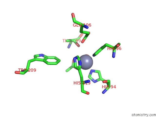
Mono view
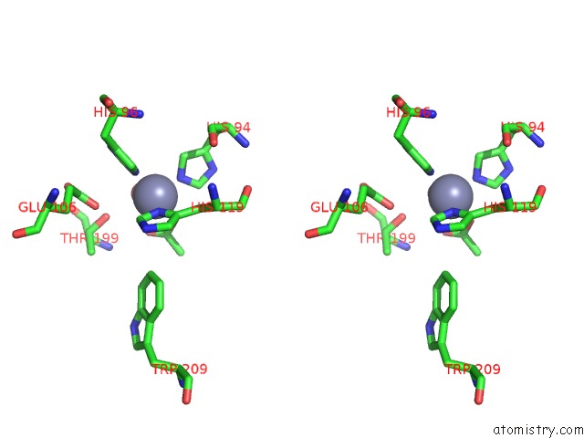
Stereo pair view
Mono view
Stereo pair view
A full contact list of Zinc with other atoms in the Zn binding
site number 2 of X-Ray Crystal Structure of Carbonic Anhydrase XII Complexed with A Theranostic Monoclonal Antibody Fragment within 5.0Å range:
|
Reference:
V.Alterio,
M.Kellner,
D.Esposito,
F.Liesche-Starnecker,
S.Bua,
C.T.Supuran,
S.M.Monti,
R.Zeidler,
G.De Simone.
Biochemical and Structural Insights Into Carbonic Anhydrase XII/FAB6A10 Complex. J.Mol.Biol. 2019.
ISSN: ESSN 1089-8638
PubMed: 31682835
DOI: 10.1016/J.JMB.2019.10.022
Page generated: Tue Oct 29 06:41:52 2024
ISSN: ESSN 1089-8638
PubMed: 31682835
DOI: 10.1016/J.JMB.2019.10.022
Last articles
Zn in 9J0NZn in 9J0O
Zn in 9J0P
Zn in 9FJX
Zn in 9EKB
Zn in 9C0F
Zn in 9CAH
Zn in 9CH0
Zn in 9CH3
Zn in 9CH1