Zinc »
PDB 6qeb-6r05 »
6qs1 »
Zinc in PDB 6qs1: Crystal Structure of Human Angiotensin-1 Converting Enzyme N-Domain in Complex with Bppb
Enzymatic activity of Crystal Structure of Human Angiotensin-1 Converting Enzyme N-Domain in Complex with Bppb
All present enzymatic activity of Crystal Structure of Human Angiotensin-1 Converting Enzyme N-Domain in Complex with Bppb:
3.4.15.1;
3.4.15.1;
Protein crystallography data
The structure of Crystal Structure of Human Angiotensin-1 Converting Enzyme N-Domain in Complex with Bppb, PDB code: 6qs1
was solved by
G.E.Cozier,
K.R.Acharya,
with X-Ray Crystallography technique. A brief refinement statistics is given in the table below:
| Resolution Low / High (Å) | 74.05 / 1.80 |
| Space group | P 1 |
| Cell size a, b, c (Å), α, β, γ (°) | 72.682, 77.070, 82.363, 88.86, 64.59, 75.03 |
| R / Rfree (%) | 20.2 / 23.2 |
Other elements in 6qs1:
The structure of Crystal Structure of Human Angiotensin-1 Converting Enzyme N-Domain in Complex with Bppb also contains other interesting chemical elements:
| Magnesium | (Mg) | 2 atoms |
| Chlorine | (Cl) | 4 atoms |
Zinc Binding Sites:
The binding sites of Zinc atom in the Crystal Structure of Human Angiotensin-1 Converting Enzyme N-Domain in Complex with Bppb
(pdb code 6qs1). This binding sites where shown within
5.0 Angstroms radius around Zinc atom.
In total 2 binding sites of Zinc where determined in the Crystal Structure of Human Angiotensin-1 Converting Enzyme N-Domain in Complex with Bppb, PDB code: 6qs1:
Jump to Zinc binding site number: 1; 2;
In total 2 binding sites of Zinc where determined in the Crystal Structure of Human Angiotensin-1 Converting Enzyme N-Domain in Complex with Bppb, PDB code: 6qs1:
Jump to Zinc binding site number: 1; 2;
Zinc binding site 1 out of 2 in 6qs1
Go back to
Zinc binding site 1 out
of 2 in the Crystal Structure of Human Angiotensin-1 Converting Enzyme N-Domain in Complex with Bppb
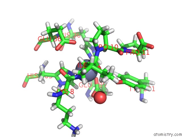
Mono view
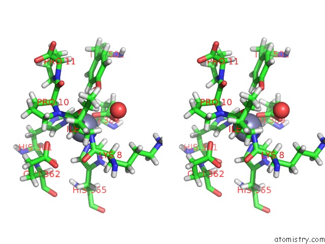
Stereo pair view
Mono view
Stereo pair view
A full contact list of Zinc with other atoms in the Zn binding
site number 1 of Crystal Structure of Human Angiotensin-1 Converting Enzyme N-Domain in Complex with Bppb within 5.0Å range:
|
Zinc binding site 2 out of 2 in 6qs1
Go back to
Zinc binding site 2 out
of 2 in the Crystal Structure of Human Angiotensin-1 Converting Enzyme N-Domain in Complex with Bppb
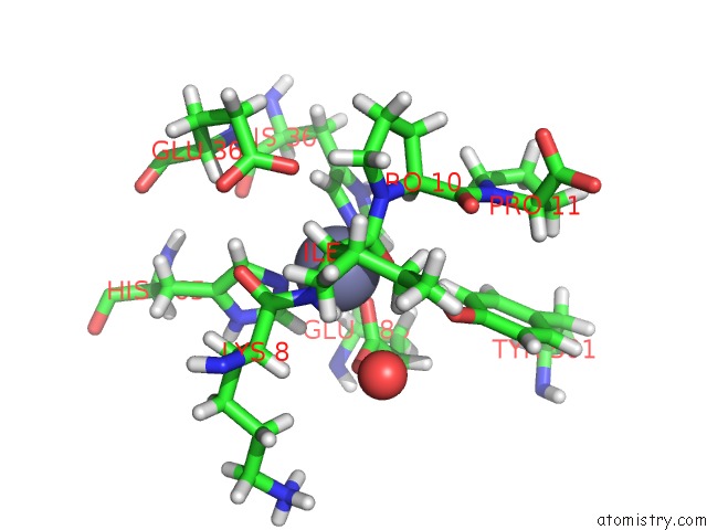
Mono view
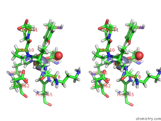
Stereo pair view
Mono view
Stereo pair view
A full contact list of Zinc with other atoms in the Zn binding
site number 2 of Crystal Structure of Human Angiotensin-1 Converting Enzyme N-Domain in Complex with Bppb within 5.0Å range:
|
Reference:
E.D.Sturrock,
L.Lubbe,
G.E.Cozier,
S.L.U.Schwager,
A.T.Arowolo,
L.B.Arendse,
E.Belcher,
K.R.Acharya.
Structural Basis For the C-Domain-Selective Angiotensin-Converting Enzyme Inhibition By Bradykinin-Potentiating Peptide B (Bppb). Biochem.J. V. 476 1553 2019.
ISSN: ESSN 1470-8728
PubMed: 31072910
DOI: 10.1042/BCJ20190290
Page generated: Tue Oct 29 05:44:25 2024
ISSN: ESSN 1470-8728
PubMed: 31072910
DOI: 10.1042/BCJ20190290
Last articles
Zn in 9J0NZn in 9J0O
Zn in 9J0P
Zn in 9FJX
Zn in 9EKB
Zn in 9C0F
Zn in 9CAH
Zn in 9CH0
Zn in 9CH3
Zn in 9CH1