Zinc »
PDB 6p6g-6pi1 »
6p9u »
Zinc in PDB 6p9u: Crystal Structure of Human Thrombin Mutant W215A
Enzymatic activity of Crystal Structure of Human Thrombin Mutant W215A
All present enzymatic activity of Crystal Structure of Human Thrombin Mutant W215A:
3.4.21.5;
3.4.21.5;
Protein crystallography data
The structure of Crystal Structure of Human Thrombin Mutant W215A, PDB code: 6p9u
was solved by
L.A.Pelc,
S.K.Koester,
Z.Chen,
E.Di Cera,
with X-Ray Crystallography technique. A brief refinement statistics is given in the table below:
| Resolution Low / High (Å) | 32.59 / 3.30 |
| Space group | P 1 2 1 |
| Cell size a, b, c (Å), α, β, γ (°) | 136.320, 44.232, 136.187, 90.00, 90.04, 90.00 |
| R / Rfree (%) | 22.5 / 30.8 |
Zinc Binding Sites:
The binding sites of Zinc atom in the Crystal Structure of Human Thrombin Mutant W215A
(pdb code 6p9u). This binding sites where shown within
5.0 Angstroms radius around Zinc atom.
In total 7 binding sites of Zinc where determined in the Crystal Structure of Human Thrombin Mutant W215A, PDB code: 6p9u:
Jump to Zinc binding site number: 1; 2; 3; 4; 5; 6; 7;
In total 7 binding sites of Zinc where determined in the Crystal Structure of Human Thrombin Mutant W215A, PDB code: 6p9u:
Jump to Zinc binding site number: 1; 2; 3; 4; 5; 6; 7;
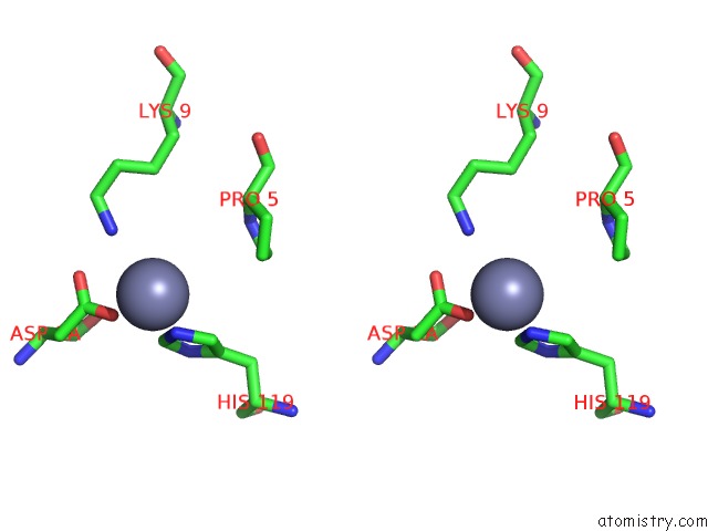
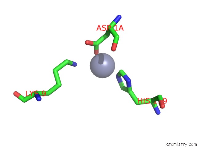
Zinc binding site 1 out of 7 in 6p9u
Go back to
Zinc binding site 1 out
of 7 in the Crystal Structure of Human Thrombin Mutant W215A

Mono view
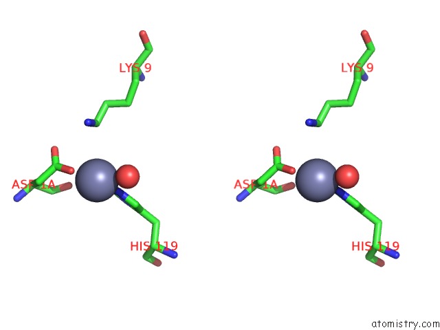
Stereo pair view
Mono view
Stereo pair view
A full contact list of Zinc with other atoms in the Zn binding
site number 1 of Crystal Structure of Human Thrombin Mutant W215A within 5.0Å range:
|
Zinc binding site 2 out of 7 in 6p9u
Go back to
Zinc binding site 2 out
of 7 in the Crystal Structure of Human Thrombin Mutant W215A
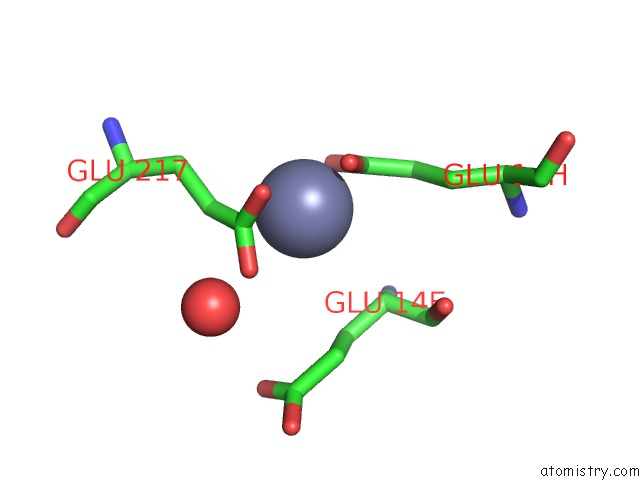
Mono view
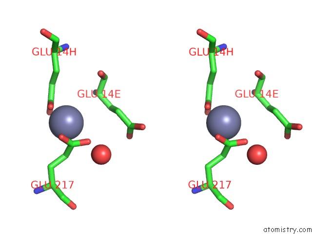
Stereo pair view
Mono view
Stereo pair view
A full contact list of Zinc with other atoms in the Zn binding
site number 2 of Crystal Structure of Human Thrombin Mutant W215A within 5.0Å range:
|
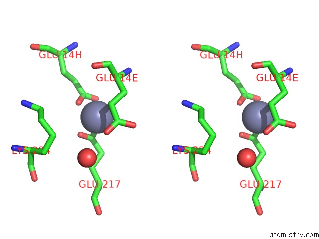
Zinc binding site 3 out of 7 in 6p9u
Go back to
Zinc binding site 3 out
of 7 in the Crystal Structure of Human Thrombin Mutant W215A

Mono view

Stereo pair view
Mono view
Stereo pair view
A full contact list of Zinc with other atoms in the Zn binding
site number 3 of Crystal Structure of Human Thrombin Mutant W215A within 5.0Å range:
|
Zinc binding site 4 out of 7 in 6p9u
Go back to
Zinc binding site 4 out
of 7 in the Crystal Structure of Human Thrombin Mutant W215A

Mono view
Stereo pair view
Mono view
Stereo pair view
A full contact list of Zinc with other atoms in the Zn binding
site number 4 of Crystal Structure of Human Thrombin Mutant W215A within 5.0Å range:
|
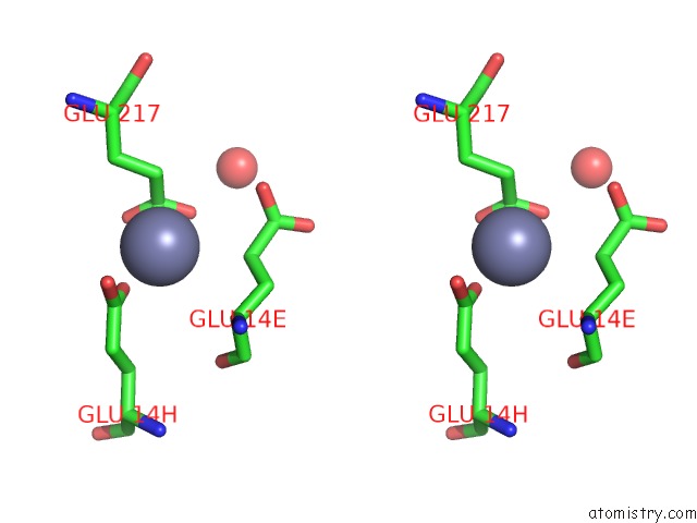
Zinc binding site 5 out of 7 in 6p9u
Go back to
Zinc binding site 5 out
of 7 in the Crystal Structure of Human Thrombin Mutant W215A

Mono view
Stereo pair view
Mono view
Stereo pair view
A full contact list of Zinc with other atoms in the Zn binding
site number 5 of Crystal Structure of Human Thrombin Mutant W215A within 5.0Å range:
|
Zinc binding site 6 out of 7 in 6p9u
Go back to
Zinc binding site 6 out
of 7 in the Crystal Structure of Human Thrombin Mutant W215A

Mono view
Stereo pair view
Mono view
Stereo pair view
A full contact list of Zinc with other atoms in the Zn binding
site number 6 of Crystal Structure of Human Thrombin Mutant W215A within 5.0Å range:
|
Zinc binding site 7 out of 7 in 6p9u
Go back to
Zinc binding site 7 out
of 7 in the Crystal Structure of Human Thrombin Mutant W215A
Mono view

Stereo pair view
Mono view
Stereo pair view
A full contact list of Zinc with other atoms in the Zn binding
site number 7 of Crystal Structure of Human Thrombin Mutant W215A within 5.0Å range:
|
Reference:
L.A.Pelc,
S.K.Koester,
Z.Chen,
N.E.Gistover,
E.Di Cera.
Residues W215, E217 and E192 Control the Allosteric E*-E Equilibrium of Thrombin. Sci Rep V. 9 12304 2019.
ISSN: ESSN 2045-2322
PubMed: 31444378
DOI: 10.1038/S41598-019-48839-1
Page generated: Tue Oct 29 04:54:18 2024
ISSN: ESSN 2045-2322
PubMed: 31444378
DOI: 10.1038/S41598-019-48839-1
Last articles
Zn in 9J0NZn in 9J0O
Zn in 9J0P
Zn in 9FJX
Zn in 9EKB
Zn in 9C0F
Zn in 9CAH
Zn in 9CH0
Zn in 9CH3
Zn in 9CH1