Zinc »
PDB 6nl8-6o5g »
6nu9 »
Zinc in PDB 6nu9: Crystal Structure of A Zinc-Binding Non-Structural Protein From the Hepatitis E Virus
Protein crystallography data
The structure of Crystal Structure of A Zinc-Binding Non-Structural Protein From the Hepatitis E Virus, PDB code: 6nu9
was solved by
A.Proudfoot,
D.Bussiere,
with X-Ray Crystallography technique. A brief refinement statistics is given in the table below:
| Resolution Low / High (Å) | 52.62 / 1.76 |
| Space group | P 65 2 2 |
| Cell size a, b, c (Å), α, β, γ (°) | 64.399, 64.399, 158.901, 90.00, 90.00, 120.00 |
| R / Rfree (%) | 23.7 / 26.9 |
Zinc Binding Sites:
The binding sites of Zinc atom in the Crystal Structure of A Zinc-Binding Non-Structural Protein From the Hepatitis E Virus
(pdb code 6nu9). This binding sites where shown within
5.0 Angstroms radius around Zinc atom.
In total only one binding site of Zinc was determined in the Crystal Structure of A Zinc-Binding Non-Structural Protein From the Hepatitis E Virus, PDB code: 6nu9:
In total only one binding site of Zinc was determined in the Crystal Structure of A Zinc-Binding Non-Structural Protein From the Hepatitis E Virus, PDB code: 6nu9:
Zinc binding site 1 out of 1 in 6nu9
Go back to
Zinc binding site 1 out
of 1 in the Crystal Structure of A Zinc-Binding Non-Structural Protein From the Hepatitis E Virus
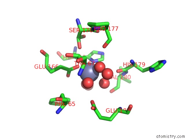
Mono view
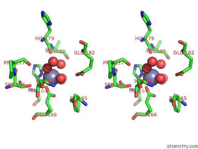
Stereo pair view
Mono view
Stereo pair view
A full contact list of Zinc with other atoms in the Zn binding
site number 1 of Crystal Structure of A Zinc-Binding Non-Structural Protein From the Hepatitis E Virus within 5.0Å range:
|
Reference:
A.Proudfoot,
A.Hyrina,
M.Holdorf,
A.O.Frank,
D.Bussiere.
First Crystal Structure of A Nonstructural Hepatitis E Viral Protein Identifies A Putative Novel Zinc-Binding Protein. J.Virol. V. 93 2019.
ISSN: ESSN 1098-5514
PubMed: 31019049
DOI: 10.1128/JVI.00170-19
Page generated: Tue Oct 29 03:59:10 2024
ISSN: ESSN 1098-5514
PubMed: 31019049
DOI: 10.1128/JVI.00170-19
Last articles
Zn in 9J0NZn in 9J0O
Zn in 9J0P
Zn in 9FJX
Zn in 9EKB
Zn in 9C0F
Zn in 9CAH
Zn in 9CH0
Zn in 9CH3
Zn in 9CH1