Zinc »
PDB 6nl8-6o5g »
6nq3 »
Zinc in PDB 6nq3: Structure of A Chromatin Complex
Protein crystallography data
The structure of Structure of A Chromatin Complex, PDB code: 6nq3
was solved by
S.Chen,
L.Jiao,
X.Liu,
with X-Ray Crystallography technique. A brief refinement statistics is given in the table below:
| Resolution Low / High (Å) | 46.60 / 2.89 |
| Space group | C 2 2 21 |
| Cell size a, b, c (Å), α, β, γ (°) | 127.710, 139.600, 268.080, 90.00, 90.00, 90.00 |
| R / Rfree (%) | 17 / 23 |
Zinc Binding Sites:
The binding sites of Zinc atom in the Structure of A Chromatin Complex
(pdb code 6nq3). This binding sites where shown within
5.0 Angstroms radius around Zinc atom.
In total 2 binding sites of Zinc where determined in the Structure of A Chromatin Complex, PDB code: 6nq3:
Jump to Zinc binding site number: 1; 2;
In total 2 binding sites of Zinc where determined in the Structure of A Chromatin Complex, PDB code: 6nq3:
Jump to Zinc binding site number: 1; 2;
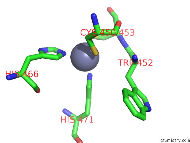
Zinc binding site 1 out of 2 in 6nq3
Go back to
Zinc binding site 1 out
of 2 in the Structure of A Chromatin Complex

Mono view
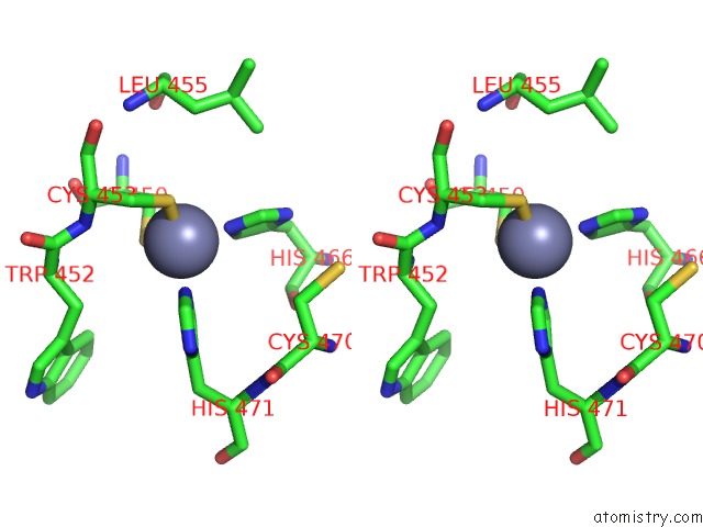
Stereo pair view
Mono view
Stereo pair view
A full contact list of Zinc with other atoms in the Zn binding
site number 1 of Structure of A Chromatin Complex within 5.0Å range:
|
Zinc binding site 2 out of 2 in 6nq3
Go back to
Zinc binding site 2 out
of 2 in the Structure of A Chromatin Complex
Mono view

Stereo pair view
Mono view
Stereo pair view
A full contact list of Zinc with other atoms in the Zn binding
site number 2 of Structure of A Chromatin Complex within 5.0Å range:
|
Reference:
S.Chen,
L.Jiao,
X.Liu,
X.Yang,
X.Liu.
A Dimeric Structural Scaffold For PRC2-Pcl Targeting to Cpg Island Chromatin. Mol.Cell 2020.
ISSN: ISSN 1097-2765
PubMed: 31959557
DOI: 10.1016/J.MOLCEL.2019.12.019
Page generated: Tue Oct 29 03:58:11 2024
ISSN: ISSN 1097-2765
PubMed: 31959557
DOI: 10.1016/J.MOLCEL.2019.12.019
Last articles
Zn in 9J0NZn in 9J0O
Zn in 9J0P
Zn in 9FJX
Zn in 9EKB
Zn in 9C0F
Zn in 9CAH
Zn in 9CH0
Zn in 9CH3
Zn in 9CH1