Zinc »
PDB 6mby-6mn4 »
6mjg »
Zinc in PDB 6mjg: Structure of Dbophma in Complex with Sah and Methylated Peptide
Protein crystallography data
The structure of Structure of Dbophma in Complex with Sah and Methylated Peptide, PDB code: 6mjg
was solved by
C.Ongpipattanakul,
S.K.Nair,
with X-Ray Crystallography technique. A brief refinement statistics is given in the table below:
| Resolution Low / High (Å) | 25.00 / 2.12 |
| Space group | C 2 2 21 |
| Cell size a, b, c (Å), α, β, γ (°) | 106.408, 107.975, 79.340, 90.00, 90.00, 90.00 |
| R / Rfree (%) | 18.6 / 22.7 |
Zinc Binding Sites:
The binding sites of Zinc atom in the Structure of Dbophma in Complex with Sah and Methylated Peptide
(pdb code 6mjg). This binding sites where shown within
5.0 Angstroms radius around Zinc atom.
In total only one binding site of Zinc was determined in the Structure of Dbophma in Complex with Sah and Methylated Peptide, PDB code: 6mjg:
In total only one binding site of Zinc was determined in the Structure of Dbophma in Complex with Sah and Methylated Peptide, PDB code: 6mjg:
Zinc binding site 1 out of 1 in 6mjg
Go back to
Zinc binding site 1 out
of 1 in the Structure of Dbophma in Complex with Sah and Methylated Peptide
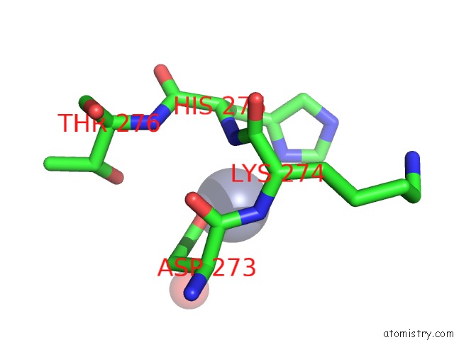
Mono view
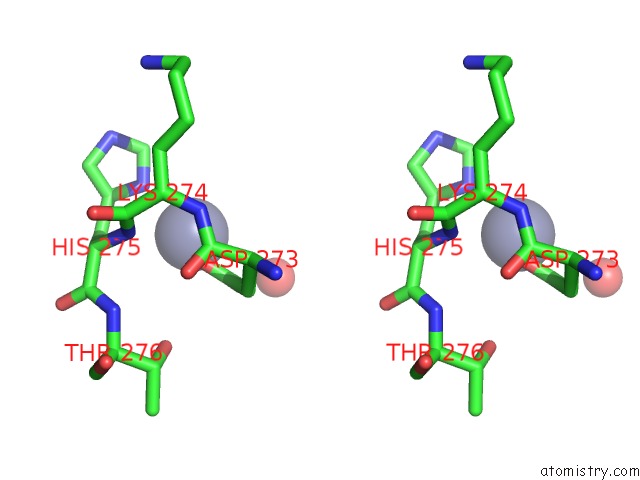
Stereo pair view
Mono view
Stereo pair view
A full contact list of Zinc with other atoms in the Zn binding
site number 1 of Structure of Dbophma in Complex with Sah and Methylated Peptide within 5.0Å range:
|
Reference:
C.Ongpipattanakul,
S.K.Nair.
Molecular Basis For Autocatalytic Backbone N-Methylation in Ripp Natural Product Biosynthesis. Acs Chem. Biol. V. 13 2989 2018.
ISSN: ESSN 1554-8937
PubMed: 30204409
DOI: 10.1021/ACSCHEMBIO.8B00668
Page generated: Tue Oct 29 03:12:53 2024
ISSN: ESSN 1554-8937
PubMed: 30204409
DOI: 10.1021/ACSCHEMBIO.8B00668
Last articles
Zn in 9J0NZn in 9J0O
Zn in 9J0P
Zn in 9FJX
Zn in 9EKB
Zn in 9C0F
Zn in 9CAH
Zn in 9CH0
Zn in 9CH3
Zn in 9CH1