Zinc »
PDB 6l72-6lj7 »
6lae »
Zinc in PDB 6lae: Crystal Structure of the Dna-Binding Domain of Human Xpa in Complex with Dna
Protein crystallography data
The structure of Crystal Structure of the Dna-Binding Domain of Human Xpa in Complex with Dna, PDB code: 6lae
was solved by
F.M.Lian,
X.Yang,
Y.L.Jiang,
F.Yang,
C.Li,
W.Yang,
C.Qian,
with X-Ray Crystallography technique. A brief refinement statistics is given in the table below:
| Resolution Low / High (Å) | 43.70 / 2.81 |
| Space group | P 31 |
| Cell size a, b, c (Å), α, β, γ (°) | 69.112, 69.112, 63.938, 90.00, 90.00, 120.00 |
| R / Rfree (%) | 21.9 / 23.7 |
Zinc Binding Sites:
The binding sites of Zinc atom in the Crystal Structure of the Dna-Binding Domain of Human Xpa in Complex with Dna
(pdb code 6lae). This binding sites where shown within
5.0 Angstroms radius around Zinc atom.
In total 2 binding sites of Zinc where determined in the Crystal Structure of the Dna-Binding Domain of Human Xpa in Complex with Dna, PDB code: 6lae:
Jump to Zinc binding site number: 1; 2;
In total 2 binding sites of Zinc where determined in the Crystal Structure of the Dna-Binding Domain of Human Xpa in Complex with Dna, PDB code: 6lae:
Jump to Zinc binding site number: 1; 2;
Zinc binding site 1 out of 2 in 6lae
Go back to
Zinc binding site 1 out
of 2 in the Crystal Structure of the Dna-Binding Domain of Human Xpa in Complex with Dna
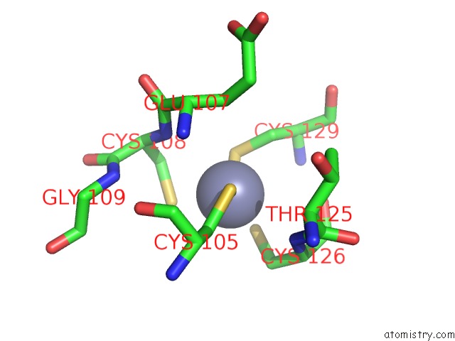
Mono view
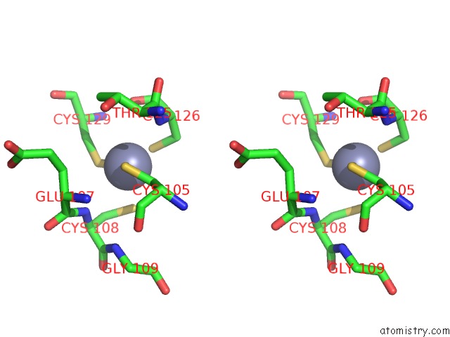
Stereo pair view
Mono view
Stereo pair view
A full contact list of Zinc with other atoms in the Zn binding
site number 1 of Crystal Structure of the Dna-Binding Domain of Human Xpa in Complex with Dna within 5.0Å range:
|
Zinc binding site 2 out of 2 in 6lae
Go back to
Zinc binding site 2 out
of 2 in the Crystal Structure of the Dna-Binding Domain of Human Xpa in Complex with Dna
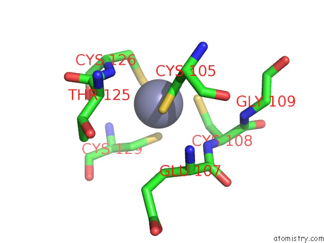
Mono view
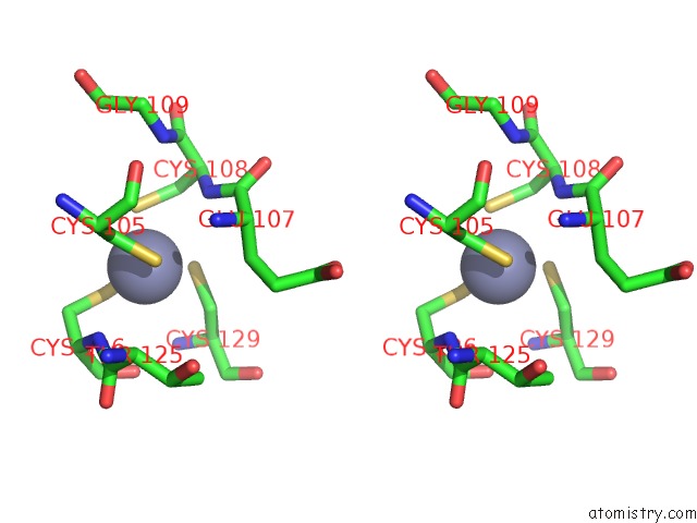
Stereo pair view
Mono view
Stereo pair view
A full contact list of Zinc with other atoms in the Zn binding
site number 2 of Crystal Structure of the Dna-Binding Domain of Human Xpa in Complex with Dna within 5.0Å range:
|
Reference:
F.M.Lian,
X.Yang,
Y.L.Jiang,
F.Yang,
C.Li,
W.Yang,
C.Qian.
New Structural Insights Into the Recognition of Undamaged Splayed-Arm Dna with A Single Pair of Non-Complementary Nucleotides By Human Nucleotide Excision Repair Protein Xpa. Int.J.Biol.Macromol. V. 148 466 2020.
ISSN: ISSN 0141-8130
PubMed: 31962067
DOI: 10.1016/J.IJBIOMAC.2020.01.169
Page generated: Tue Oct 29 02:27:40 2024
ISSN: ISSN 0141-8130
PubMed: 31962067
DOI: 10.1016/J.IJBIOMAC.2020.01.169
Last articles
Zn in 9J0NZn in 9J0O
Zn in 9J0P
Zn in 9FJX
Zn in 9EKB
Zn in 9C0F
Zn in 9CAH
Zn in 9CH0
Zn in 9CH3
Zn in 9CH1