Zinc »
PDB 6jqx-6kal »
6jzv »
Zinc in PDB 6jzv: Crystal Structure of Sufu From Bacillus Subtilis
Protein crystallography data
The structure of Crystal Structure of Sufu From Bacillus Subtilis, PDB code: 6jzv
was solved by
T.Fujishiro,
Y.Takahashi,
with X-Ray Crystallography technique. A brief refinement statistics is given in the table below:
| Resolution Low / High (Å) | 49.41 / 2.00 |
| Space group | P 1 |
| Cell size a, b, c (Å), α, β, γ (°) | 35.320, 54.450, 70.380, 68.99, 75.86, 72.54 |
| R / Rfree (%) | 21.2 / 24.2 |
Zinc Binding Sites:
The binding sites of Zinc atom in the Crystal Structure of Sufu From Bacillus Subtilis
(pdb code 6jzv). This binding sites where shown within
5.0 Angstroms radius around Zinc atom.
In total 4 binding sites of Zinc where determined in the Crystal Structure of Sufu From Bacillus Subtilis, PDB code: 6jzv:
Jump to Zinc binding site number: 1; 2; 3; 4;
In total 4 binding sites of Zinc where determined in the Crystal Structure of Sufu From Bacillus Subtilis, PDB code: 6jzv:
Jump to Zinc binding site number: 1; 2; 3; 4;
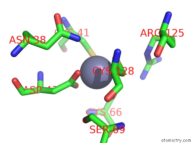
Zinc binding site 1 out of 4 in 6jzv
Go back to
Zinc binding site 1 out
of 4 in the Crystal Structure of Sufu From Bacillus Subtilis
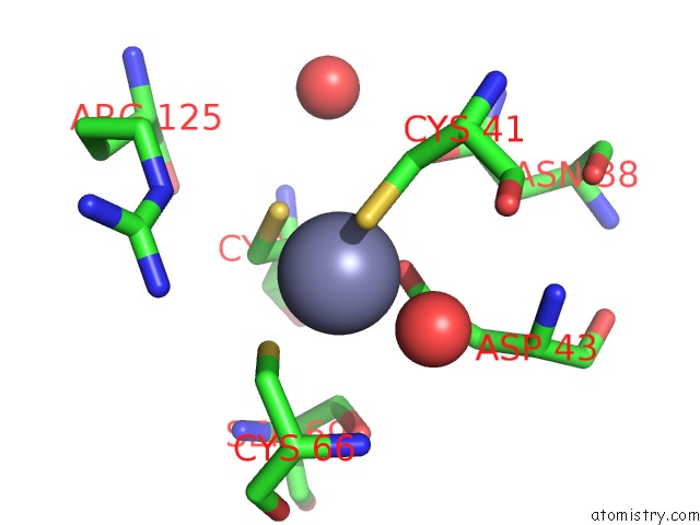
Mono view
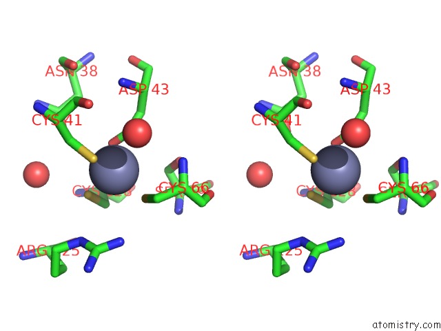
Stereo pair view
Mono view
Stereo pair view
A full contact list of Zinc with other atoms in the Zn binding
site number 1 of Crystal Structure of Sufu From Bacillus Subtilis within 5.0Å range:
|
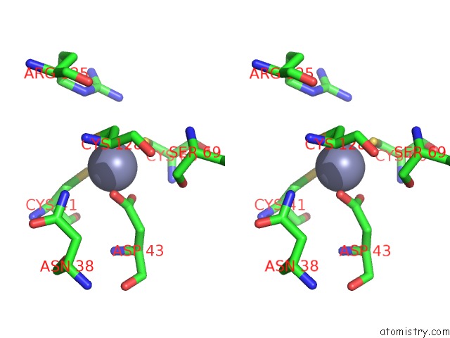
Zinc binding site 2 out of 4 in 6jzv
Go back to
Zinc binding site 2 out
of 4 in the Crystal Structure of Sufu From Bacillus Subtilis
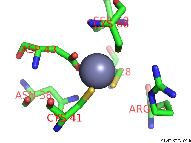
Mono view
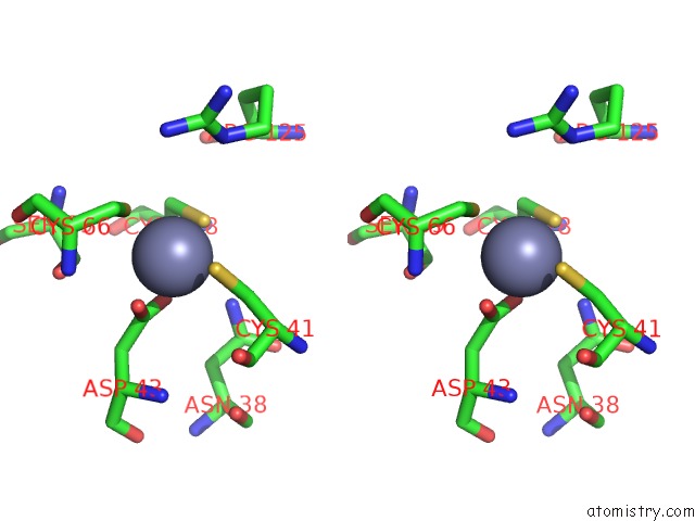
Stereo pair view
Mono view
Stereo pair view
A full contact list of Zinc with other atoms in the Zn binding
site number 2 of Crystal Structure of Sufu From Bacillus Subtilis within 5.0Å range:
|
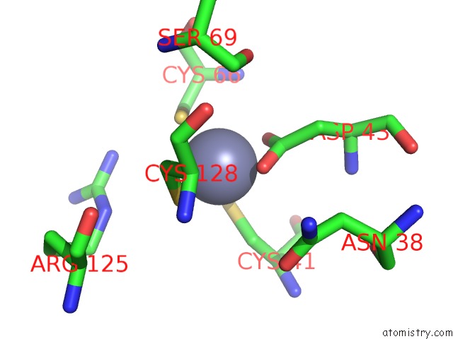
Zinc binding site 3 out of 4 in 6jzv
Go back to
Zinc binding site 3 out
of 4 in the Crystal Structure of Sufu From Bacillus Subtilis
Mono view
Stereo pair view
Mono view
Stereo pair view
A full contact list of Zinc with other atoms in the Zn binding
site number 3 of Crystal Structure of Sufu From Bacillus Subtilis within 5.0Å range:
|
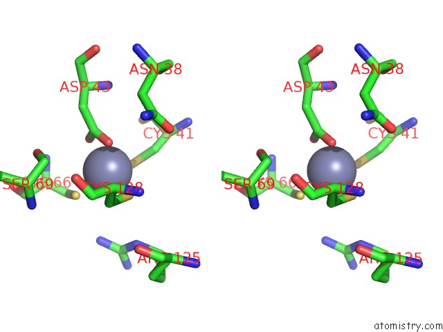
Zinc binding site 4 out of 4 in 6jzv
Go back to
Zinc binding site 4 out
of 4 in the Crystal Structure of Sufu From Bacillus Subtilis
Mono view
Stereo pair view
Mono view
Stereo pair view
A full contact list of Zinc with other atoms in the Zn binding
site number 4 of Crystal Structure of Sufu From Bacillus Subtilis within 5.0Å range:
|
Reference:
T.Fujishiro,
T.Terahata,
Y.Shimada,
Y.Takahashi.
Zinc-Persulfide Complex For Sulfur Mobilization By Sufu in Suf-Like Machinery For Fe-S Cluster Biosynthesis To Be Published.
Page generated: Tue Oct 29 01:27:28 2024
Last articles
Zn in 9J0NZn in 9J0O
Zn in 9J0P
Zn in 9FJX
Zn in 9EKB
Zn in 9C0F
Zn in 9CAH
Zn in 9CH0
Zn in 9CH3
Zn in 9CH1