Zinc »
PDB 6jet-6jqw »
6jf6 »
Zinc in PDB 6jf6: Met-Ala-Ser Bound Crystal Structure of Class I Type B Peptide Deformylase From Acinetobacter Baumannii
Enzymatic activity of Met-Ala-Ser Bound Crystal Structure of Class I Type B Peptide Deformylase From Acinetobacter Baumannii
All present enzymatic activity of Met-Ala-Ser Bound Crystal Structure of Class I Type B Peptide Deformylase From Acinetobacter Baumannii:
3.5.1.88;
3.5.1.88;
Protein crystallography data
The structure of Met-Ala-Ser Bound Crystal Structure of Class I Type B Peptide Deformylase From Acinetobacter Baumannii, PDB code: 6jf6
was solved by
K.H.Jung,
T.H.Ho,
I.H.Lee,
L.W.Kang,
with X-Ray Crystallography technique. A brief refinement statistics is given in the table below:
| Resolution Low / High (Å) | 49.99 / 2.35 |
| Space group | P 1 21 1 |
| Cell size a, b, c (Å), α, β, γ (°) | 71.370, 39.347, 110.353, 90.00, 89.98, 90.00 |
| R / Rfree (%) | 21 / 29.2 |
Zinc Binding Sites:
The binding sites of Zinc atom in the Met-Ala-Ser Bound Crystal Structure of Class I Type B Peptide Deformylase From Acinetobacter Baumannii
(pdb code 6jf6). This binding sites where shown within
5.0 Angstroms radius around Zinc atom.
In total 4 binding sites of Zinc where determined in the Met-Ala-Ser Bound Crystal Structure of Class I Type B Peptide Deformylase From Acinetobacter Baumannii, PDB code: 6jf6:
Jump to Zinc binding site number: 1; 2; 3; 4;
In total 4 binding sites of Zinc where determined in the Met-Ala-Ser Bound Crystal Structure of Class I Type B Peptide Deformylase From Acinetobacter Baumannii, PDB code: 6jf6:
Jump to Zinc binding site number: 1; 2; 3; 4;
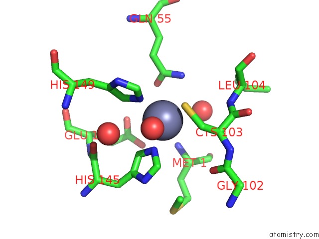
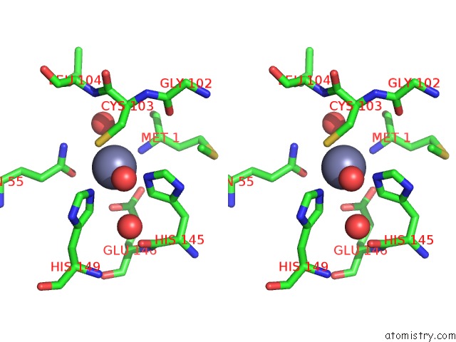
Zinc binding site 1 out of 4 in 6jf6
Go back to
Zinc binding site 1 out
of 4 in the Met-Ala-Ser Bound Crystal Structure of Class I Type B Peptide Deformylase From Acinetobacter Baumannii
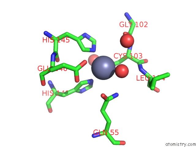
Mono view
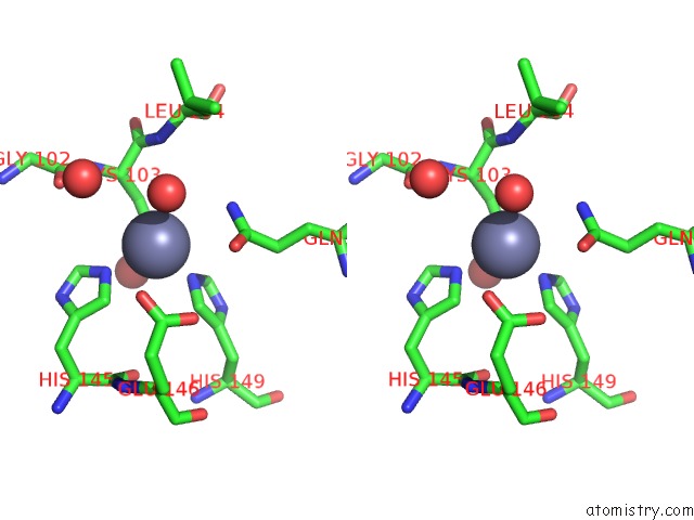
Stereo pair view
Mono view
Stereo pair view
A full contact list of Zinc with other atoms in the Zn binding
site number 1 of Met-Ala-Ser Bound Crystal Structure of Class I Type B Peptide Deformylase From Acinetobacter Baumannii within 5.0Å range:
|
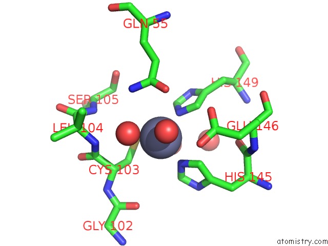
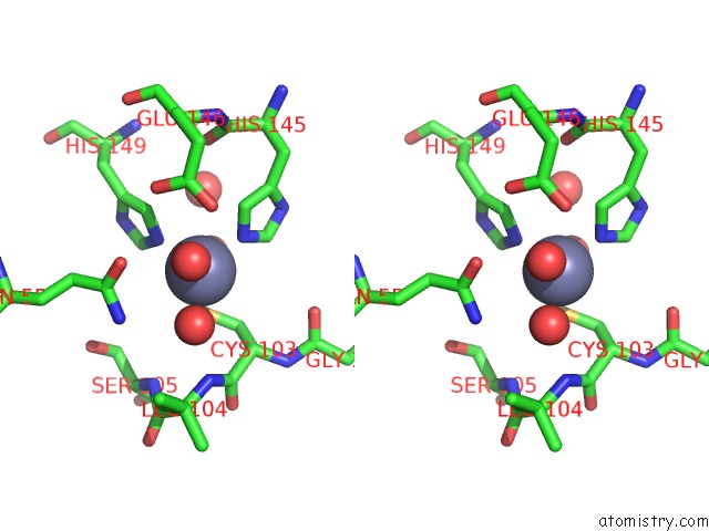
Zinc binding site 2 out of 4 in 6jf6
Go back to
Zinc binding site 2 out
of 4 in the Met-Ala-Ser Bound Crystal Structure of Class I Type B Peptide Deformylase From Acinetobacter Baumannii
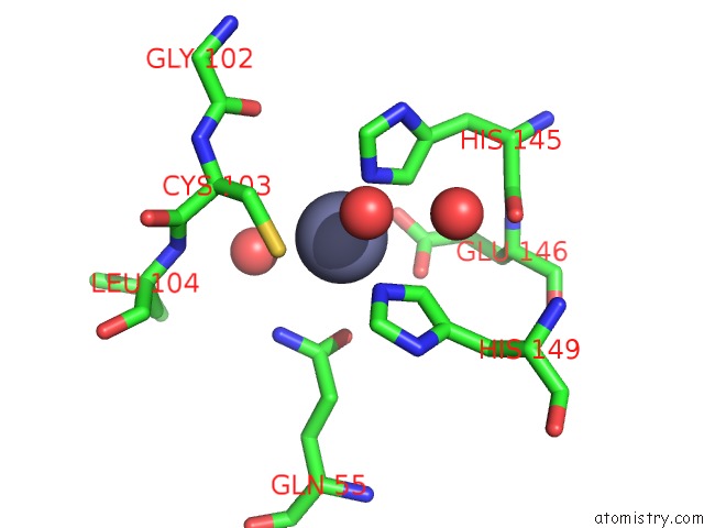
Mono view
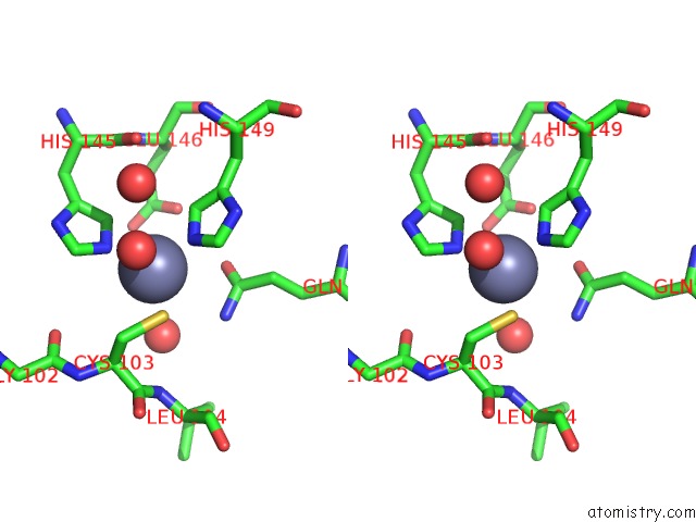
Stereo pair view
Mono view
Stereo pair view
A full contact list of Zinc with other atoms in the Zn binding
site number 2 of Met-Ala-Ser Bound Crystal Structure of Class I Type B Peptide Deformylase From Acinetobacter Baumannii within 5.0Å range:
|
Zinc binding site 3 out of 4 in 6jf6
Go back to
Zinc binding site 3 out
of 4 in the Met-Ala-Ser Bound Crystal Structure of Class I Type B Peptide Deformylase From Acinetobacter Baumannii
Mono view
Stereo pair view
Mono view
Stereo pair view
A full contact list of Zinc with other atoms in the Zn binding
site number 3 of Met-Ala-Ser Bound Crystal Structure of Class I Type B Peptide Deformylase From Acinetobacter Baumannii within 5.0Å range:
|
Zinc binding site 4 out of 4 in 6jf6
Go back to
Zinc binding site 4 out
of 4 in the Met-Ala-Ser Bound Crystal Structure of Class I Type B Peptide Deformylase From Acinetobacter Baumannii
Mono view
Stereo pair view
Mono view
Stereo pair view
A full contact list of Zinc with other atoms in the Zn binding
site number 4 of Met-Ala-Ser Bound Crystal Structure of Class I Type B Peptide Deformylase From Acinetobacter Baumannii within 5.0Å range:
|
Reference:
K.H.Jung,
T.H.Ho,
I.H.Lee,
L.W.Kang.
Met-Ala-Ser Bound Crystal Structure of Class I Type B Peptide Deformylase From Acinetobacter Baumannii To Be Published.
Page generated: Tue Oct 29 01:07:12 2024
Last articles
Zn in 9J0NZn in 9J0O
Zn in 9J0P
Zn in 9FJX
Zn in 9EKB
Zn in 9C0F
Zn in 9CAH
Zn in 9CH0
Zn in 9CH3
Zn in 9CH1