Zinc »
PDB 6i0w-6iiw »
6ig7 »
Zinc in PDB 6ig7: Crystal Structure of Thermolysin Delivered in Polyacrylamide Using X- Ray Free Electron Laser
Enzymatic activity of Crystal Structure of Thermolysin Delivered in Polyacrylamide Using X- Ray Free Electron Laser
All present enzymatic activity of Crystal Structure of Thermolysin Delivered in Polyacrylamide Using X- Ray Free Electron Laser:
3.4.24.27;
3.4.24.27;
Protein crystallography data
The structure of Crystal Structure of Thermolysin Delivered in Polyacrylamide Using X- Ray Free Electron Laser, PDB code: 6ig7
was solved by
K.H.Nam,
with X-Ray Crystallography technique. A brief refinement statistics is given in the table below:
| Resolution Low / High (Å) | 40.12 / 1.80 |
| Space group | P 61 2 2 |
| Cell size a, b, c (Å), α, β, γ (°) | 92.663, 92.663, 128.591, 90.00, 90.00, 120.00 |
| R / Rfree (%) | 17.3 / 20.2 |
Other elements in 6ig7:
The structure of Crystal Structure of Thermolysin Delivered in Polyacrylamide Using X- Ray Free Electron Laser also contains other interesting chemical elements:
| Calcium | (Ca) | 4 atoms |
Zinc Binding Sites:
The binding sites of Zinc atom in the Crystal Structure of Thermolysin Delivered in Polyacrylamide Using X- Ray Free Electron Laser
(pdb code 6ig7). This binding sites where shown within
5.0 Angstroms radius around Zinc atom.
In total only one binding site of Zinc was determined in the Crystal Structure of Thermolysin Delivered in Polyacrylamide Using X- Ray Free Electron Laser, PDB code: 6ig7:
In total only one binding site of Zinc was determined in the Crystal Structure of Thermolysin Delivered in Polyacrylamide Using X- Ray Free Electron Laser, PDB code: 6ig7:
Zinc binding site 1 out of 1 in 6ig7
Go back to
Zinc binding site 1 out
of 1 in the Crystal Structure of Thermolysin Delivered in Polyacrylamide Using X- Ray Free Electron Laser
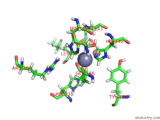
Mono view
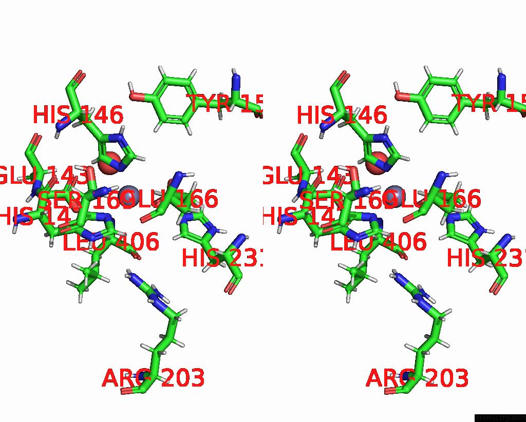
Stereo pair view
Mono view
Stereo pair view
A full contact list of Zinc with other atoms in the Zn binding
site number 1 of Crystal Structure of Thermolysin Delivered in Polyacrylamide Using X- Ray Free Electron Laser within 5.0Å range:
|
Reference:
J.Park,
S.Park,
J.Kim,
G.Park,
Y.Cho,
K.H.Nam.
Polyacrylamide Injection Matrix For Serial Femtosecond Crystallography. Sci Rep V. 9 2525 2019.
ISSN: ESSN 2045-2322
PubMed: 30792457
DOI: 10.1038/S41598-019-39020-9
Page generated: Mon Oct 28 23:49:57 2024
ISSN: ESSN 2045-2322
PubMed: 30792457
DOI: 10.1038/S41598-019-39020-9
Last articles
Zn in 9J0NZn in 9J0O
Zn in 9J0P
Zn in 9FJX
Zn in 9EKB
Zn in 9C0F
Zn in 9CAH
Zn in 9CH0
Zn in 9CH3
Zn in 9CH1