Zinc »
PDB 6i0w-6iiw »
6ifn »
Zinc in PDB 6ifn: Crystal Structure of Type III-A Crispr Csm Complex
Protein crystallography data
The structure of Crystal Structure of Type III-A Crispr Csm Complex, PDB code: 6ifn
was solved by
L.You,
J.Wang,
Y.Wang,
with X-Ray Crystallography technique. A brief refinement statistics is given in the table below:
| Resolution Low / High (Å) | 48.68 / 2.90 |
| Space group | P 1 21 1 |
| Cell size a, b, c (Å), α, β, γ (°) | 123.543, 82.334, 161.358, 90.00, 99.32, 90.00 |
| R / Rfree (%) | 20.1 / 24.4 |
Other elements in 6ifn:
The structure of Crystal Structure of Type III-A Crispr Csm Complex also contains other interesting chemical elements:
| Manganese | (Mn) | 1 atom |
Zinc Binding Sites:
The binding sites of Zinc atom in the Crystal Structure of Type III-A Crispr Csm Complex
(pdb code 6ifn). This binding sites where shown within
5.0 Angstroms radius around Zinc atom.
In total only one binding site of Zinc was determined in the Crystal Structure of Type III-A Crispr Csm Complex, PDB code: 6ifn:
In total only one binding site of Zinc was determined in the Crystal Structure of Type III-A Crispr Csm Complex, PDB code: 6ifn:
Zinc binding site 1 out of 1 in 6ifn
Go back to
Zinc binding site 1 out
of 1 in the Crystal Structure of Type III-A Crispr Csm Complex
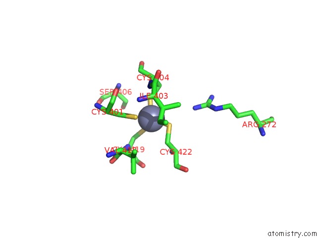
Mono view
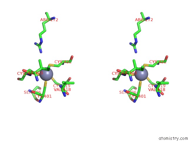
Stereo pair view
Mono view
Stereo pair view
A full contact list of Zinc with other atoms in the Zn binding
site number 1 of Crystal Structure of Type III-A Crispr Csm Complex within 5.0Å range:
|
Reference:
L.You,
J.Ma,
J.Wang,
D.Artamonova,
M.Wang,
L.Liu,
H.Xiang,
K.Severinov,
X.Zhang,
Y.Wang.
Structure Studies of the Crispr-Csm Complex Reveal Mechanism of Co-Transcriptional Interference Cell V. 176 239 2019.
ISSN: ISSN 1097-4172
PubMed: 30503210
DOI: 10.1016/J.CELL.2018.10.052
Page generated: Mon Oct 28 23:49:08 2024
ISSN: ISSN 1097-4172
PubMed: 30503210
DOI: 10.1016/J.CELL.2018.10.052
Last articles
Zn in 9J0NZn in 9J0O
Zn in 9J0P
Zn in 9FJX
Zn in 9EKB
Zn in 9C0F
Zn in 9CAH
Zn in 9CH0
Zn in 9CH3
Zn in 9CH1