Zinc »
PDB 6g3o-6gcn »
6gbx »
Zinc in PDB 6gbx: Crystal Structure of Human Glutaminyl Cyclase Variant Y115E-Y117E in Complex with SEN177
Enzymatic activity of Crystal Structure of Human Glutaminyl Cyclase Variant Y115E-Y117E in Complex with SEN177
All present enzymatic activity of Crystal Structure of Human Glutaminyl Cyclase Variant Y115E-Y117E in Complex with SEN177:
2.3.2.5;
2.3.2.5;
Protein crystallography data
The structure of Crystal Structure of Human Glutaminyl Cyclase Variant Y115E-Y117E in Complex with SEN177, PDB code: 6gbx
was solved by
C.Pozzi,
F.Di Pisa,
M.Benvenuti,
S.Mangani,
with X-Ray Crystallography technique. A brief refinement statistics is given in the table below:
| Resolution Low / High (Å) | 95.23 / 1.72 |
| Space group | C 1 2 1 |
| Cell size a, b, c (Å), α, β, γ (°) | 86.140, 149.810, 95.980, 90.00, 97.18, 90.00 |
| R / Rfree (%) | 15.8 / 18.3 |
Other elements in 6gbx:
The structure of Crystal Structure of Human Glutaminyl Cyclase Variant Y115E-Y117E in Complex with SEN177 also contains other interesting chemical elements:
| Fluorine | (F) | 3 atoms |
Zinc Binding Sites:
The binding sites of Zinc atom in the Crystal Structure of Human Glutaminyl Cyclase Variant Y115E-Y117E in Complex with SEN177
(pdb code 6gbx). This binding sites where shown within
5.0 Angstroms radius around Zinc atom.
In total 3 binding sites of Zinc where determined in the Crystal Structure of Human Glutaminyl Cyclase Variant Y115E-Y117E in Complex with SEN177, PDB code: 6gbx:
Jump to Zinc binding site number: 1; 2; 3;
In total 3 binding sites of Zinc where determined in the Crystal Structure of Human Glutaminyl Cyclase Variant Y115E-Y117E in Complex with SEN177, PDB code: 6gbx:
Jump to Zinc binding site number: 1; 2; 3;
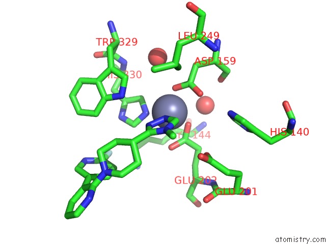
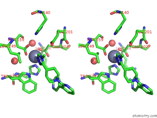
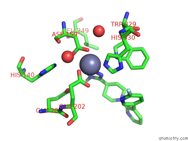
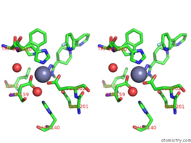
Zinc binding site 1 out of 3 in 6gbx
Go back to
Zinc binding site 1 out
of 3 in the Crystal Structure of Human Glutaminyl Cyclase Variant Y115E-Y117E in Complex with SEN177
Mono view
Stereo pair view
Mono view
Stereo pair view
A full contact list of Zinc with other atoms in the Zn binding
site number 1 of Crystal Structure of Human Glutaminyl Cyclase Variant Y115E-Y117E in Complex with SEN177 within 5.0Å range:
|
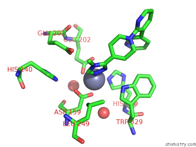
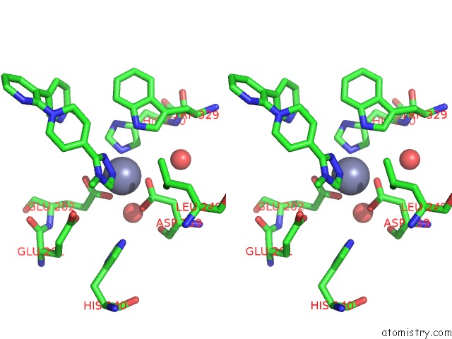
Zinc binding site 2 out of 3 in 6gbx
Go back to
Zinc binding site 2 out
of 3 in the Crystal Structure of Human Glutaminyl Cyclase Variant Y115E-Y117E in Complex with SEN177
Mono view
Stereo pair view
Mono view
Stereo pair view
A full contact list of Zinc with other atoms in the Zn binding
site number 2 of Crystal Structure of Human Glutaminyl Cyclase Variant Y115E-Y117E in Complex with SEN177 within 5.0Å range:
|
Zinc binding site 3 out of 3 in 6gbx
Go back to
Zinc binding site 3 out
of 3 in the Crystal Structure of Human Glutaminyl Cyclase Variant Y115E-Y117E in Complex with SEN177
Mono view
Stereo pair view
Mono view
Stereo pair view
A full contact list of Zinc with other atoms in the Zn binding
site number 3 of Crystal Structure of Human Glutaminyl Cyclase Variant Y115E-Y117E in Complex with SEN177 within 5.0Å range:
|
Reference:
C.Pozzi,
F.Di Pisa,
M.Benvenuti,
S.Mangani.
The Structure of the Human Glutaminyl Cyclase-SEN177 Complex Indicates Routes For Developing New Potent Inhibitors As Possible Agents For the Treatment of Neurological Disorders. J. Biol. Inorg. Chem. V. 23 1219 2018.
ISSN: ESSN 1432-1327
PubMed: 30132075
DOI: 10.1007/S00775-018-1605-1
Page generated: Mon Oct 28 21:43:53 2024
ISSN: ESSN 1432-1327
PubMed: 30132075
DOI: 10.1007/S00775-018-1605-1
Last articles
Zn in 9J0NZn in 9J0O
Zn in 9J0P
Zn in 9FJX
Zn in 9EKB
Zn in 9C0F
Zn in 9CAH
Zn in 9CH0
Zn in 9CH3
Zn in 9CH1