Zinc »
PDB 6g3o-6gcn »
6gat »
Zinc in PDB 6gat: Solution uc(Nmr) Structure of the L22V Mutant Dna Binding Domain of Area Complexed to A 13 Bp Dna Containing A Tgata Site, Regularized Mean Structure
Zinc Binding Sites:
The binding sites of Zinc atom in the Solution uc(Nmr) Structure of the L22V Mutant Dna Binding Domain of Area Complexed to A 13 Bp Dna Containing A Tgata Site, Regularized Mean Structure
(pdb code 6gat). This binding sites where shown within
5.0 Angstroms radius around Zinc atom.
In total only one binding site of Zinc was determined in the Solution uc(Nmr) Structure of the L22V Mutant Dna Binding Domain of Area Complexed to A 13 Bp Dna Containing A Tgata Site, Regularized Mean Structure, PDB code: 6gat:
In total only one binding site of Zinc was determined in the Solution uc(Nmr) Structure of the L22V Mutant Dna Binding Domain of Area Complexed to A 13 Bp Dna Containing A Tgata Site, Regularized Mean Structure, PDB code: 6gat:
Zinc binding site 1 out of 1 in 6gat
Go back to
Zinc binding site 1 out
of 1 in the Solution uc(Nmr) Structure of the L22V Mutant Dna Binding Domain of Area Complexed to A 13 Bp Dna Containing A Tgata Site, Regularized Mean Structure
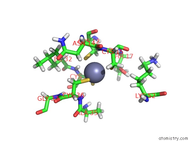
Mono view
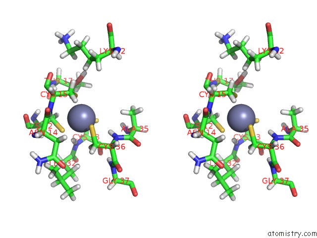
Stereo pair view
Mono view
Stereo pair view
A full contact list of Zinc with other atoms in the Zn binding
site number 1 of Solution uc(Nmr) Structure of the L22V Mutant Dna Binding Domain of Area Complexed to A 13 Bp Dna Containing A Tgata Site, Regularized Mean Structure within 5.0Å range:
|
Reference:
M.R.Starich,
M.Wikstrom,
S.Schumacher,
H.N.Arst Jr.,
A.M.Gronenborn,
G.M.Clore.
The Solution Structure of the LEU22-->Val Mutant Area Dna Binding Domain Complexed with A Tgatag Core Element Defines A Role For Hydrophobic Packing in the Determination of Specificity. J.Mol.Biol. V. 277 621 1998.
ISSN: ISSN 0022-2836
PubMed: 9533884
DOI: 10.1006/JMBI.1997.1626
Page generated: Mon Oct 28 21:42:57 2024
ISSN: ISSN 0022-2836
PubMed: 9533884
DOI: 10.1006/JMBI.1997.1626
Last articles
Zn in 9J0NZn in 9J0O
Zn in 9J0P
Zn in 9FJX
Zn in 9EKB
Zn in 9C0F
Zn in 9CAH
Zn in 9CH0
Zn in 9CH3
Zn in 9CH1