Zinc »
PDB 6bh7-6bsm »
6bms »
Zinc in PDB 6bms: Palmitoyltransferase Structure
Enzymatic activity of Palmitoyltransferase Structure
All present enzymatic activity of Palmitoyltransferase Structure:
2.3.1.225;
2.3.1.225;
Protein crystallography data
The structure of Palmitoyltransferase Structure, PDB code: 6bms
was solved by
P.Kumar,
K.Rajashankar,
with X-Ray Crystallography technique. A brief refinement statistics is given in the table below:
| Resolution Low / High (Å) | 37.21 / 2.44 |
| Space group | P 42 21 2 |
| Cell size a, b, c (Å), α, β, γ (°) | 186.402, 186.402, 90.177, 90.00, 90.00, 90.00 |
| R / Rfree (%) | 23.3 / 26.1 |
Zinc Binding Sites:
The binding sites of Zinc atom in the Palmitoyltransferase Structure
(pdb code 6bms). This binding sites where shown within
5.0 Angstroms radius around Zinc atom.
In total 4 binding sites of Zinc where determined in the Palmitoyltransferase Structure, PDB code: 6bms:
Jump to Zinc binding site number: 1; 2; 3; 4;
In total 4 binding sites of Zinc where determined in the Palmitoyltransferase Structure, PDB code: 6bms:
Jump to Zinc binding site number: 1; 2; 3; 4;
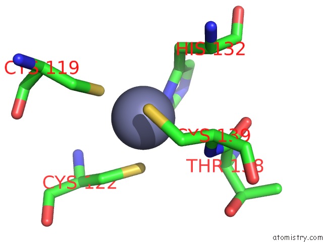
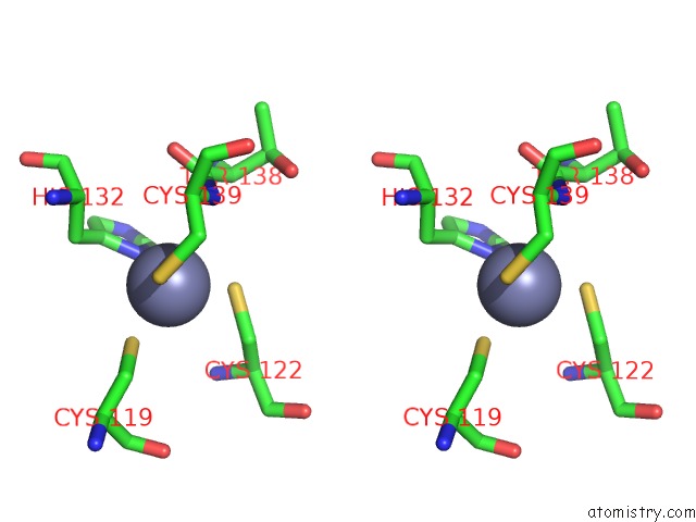
Zinc binding site 1 out of 4 in 6bms
Go back to
Zinc binding site 1 out
of 4 in the Palmitoyltransferase Structure
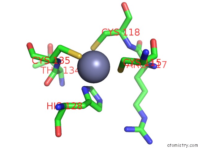
Mono view
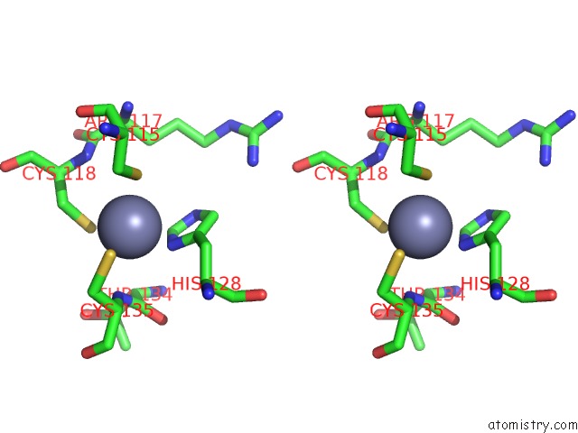
Stereo pair view
Mono view
Stereo pair view
A full contact list of Zinc with other atoms in the Zn binding
site number 1 of Palmitoyltransferase Structure within 5.0Å range:
|
Zinc binding site 2 out of 4 in 6bms
Go back to
Zinc binding site 2 out
of 4 in the Palmitoyltransferase Structure
Mono view
Stereo pair view
Mono view
Stereo pair view
A full contact list of Zinc with other atoms in the Zn binding
site number 2 of Palmitoyltransferase Structure within 5.0Å range:
|
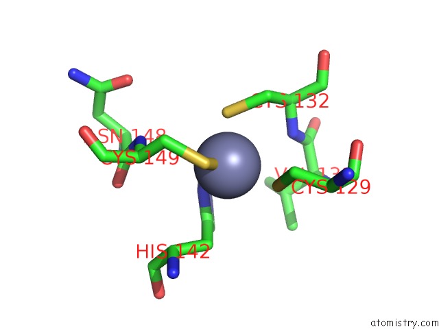
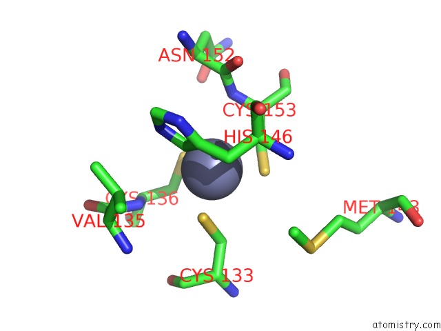
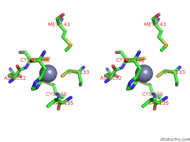
Zinc binding site 3 out of 4 in 6bms
Go back to
Zinc binding site 3 out
of 4 in the Palmitoyltransferase Structure
Mono view
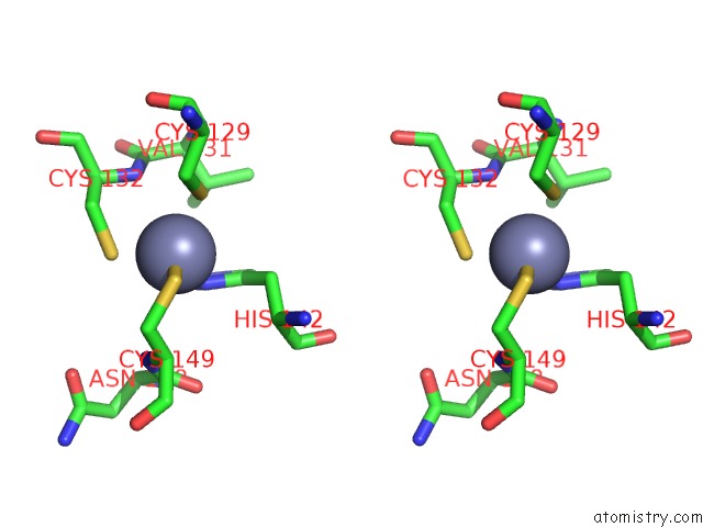
Stereo pair view
Mono view
Stereo pair view
A full contact list of Zinc with other atoms in the Zn binding
site number 3 of Palmitoyltransferase Structure within 5.0Å range:
|
Zinc binding site 4 out of 4 in 6bms
Go back to
Zinc binding site 4 out
of 4 in the Palmitoyltransferase Structure
Mono view
Stereo pair view
Mono view
Stereo pair view
A full contact list of Zinc with other atoms in the Zn binding
site number 4 of Palmitoyltransferase Structure within 5.0Å range:
|
Reference:
M.S.Rana,
P.Kumar,
C.J.Lee,
R.Verardi,
K.R.Rajashankar,
A.Banerjee.
Fatty Acyl Recognition and Transfer By An Integral Membranes-Acyltransferase. Science V. 359 2018.
ISSN: ESSN 1095-9203
PubMed: 29326245
DOI: 10.1126/SCIENCE.AAO6326
Page generated: Mon Oct 28 18:08:31 2024
ISSN: ESSN 1095-9203
PubMed: 29326245
DOI: 10.1126/SCIENCE.AAO6326
Last articles
Zn in 9J0NZn in 9J0O
Zn in 9J0P
Zn in 9FJX
Zn in 9EKB
Zn in 9C0F
Zn in 9CAH
Zn in 9CH0
Zn in 9CH3
Zn in 9CH1