Zinc »
PDB 5znl-6a3z »
5zxw »
Zinc in PDB 5zxw: Crystal Structure of Human Carbonic Anhydrase II Crystallized By Ammonium Sulfate
Enzymatic activity of Crystal Structure of Human Carbonic Anhydrase II Crystallized By Ammonium Sulfate
All present enzymatic activity of Crystal Structure of Human Carbonic Anhydrase II Crystallized By Ammonium Sulfate:
4.2.1.1;
4.2.1.1;
Protein crystallography data
The structure of Crystal Structure of Human Carbonic Anhydrase II Crystallized By Ammonium Sulfate, PDB code: 5zxw
was solved by
M.Kitahara,
S.Fudo,
T.Yoneda,
M.Nukaga,
T.Hoshino,
with X-Ray Crystallography technique. A brief refinement statistics is given in the table below:
| Resolution Low / High (Å) | 34.97 / 1.32 |
| Space group | P 1 21 1 |
| Cell size a, b, c (Å), α, β, γ (°) | 42.102, 41.323, 72.130, 90.00, 104.19, 90.00 |
| R / Rfree (%) | 17.1 / 19.8 |
Zinc Binding Sites:
The binding sites of Zinc atom in the Crystal Structure of Human Carbonic Anhydrase II Crystallized By Ammonium Sulfate
(pdb code 5zxw). This binding sites where shown within
5.0 Angstroms radius around Zinc atom.
In total only one binding site of Zinc was determined in the Crystal Structure of Human Carbonic Anhydrase II Crystallized By Ammonium Sulfate, PDB code: 5zxw:
In total only one binding site of Zinc was determined in the Crystal Structure of Human Carbonic Anhydrase II Crystallized By Ammonium Sulfate, PDB code: 5zxw:
Zinc binding site 1 out of 1 in 5zxw
Go back to
Zinc binding site 1 out
of 1 in the Crystal Structure of Human Carbonic Anhydrase II Crystallized By Ammonium Sulfate
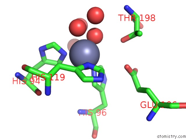
Mono view
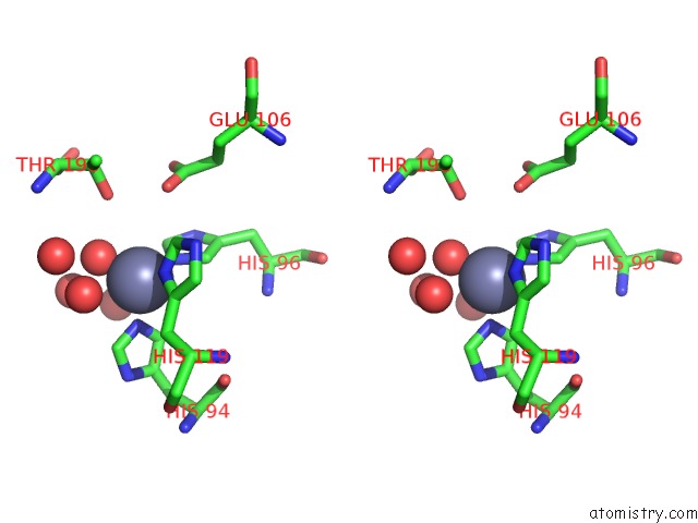
Stereo pair view
Mono view
Stereo pair view
A full contact list of Zinc with other atoms in the Zn binding
site number 1 of Crystal Structure of Human Carbonic Anhydrase II Crystallized By Ammonium Sulfate within 5.0Å range:
|
Reference:
M.Kitahara,
S.Fudo,
T.Yoneda,
M.Nukaga,
T.Hoshino.
Anisotropic Distribution of Ammonium Sulfate Ions in Protein Crystallization Cryst.Growth Des. V. 19 6004 2019.
ISSN: ESSN 1528-7505
DOI: 10.1021/ACS.CGD.9B00256
Page generated: Mon Oct 28 17:12:19 2024
ISSN: ESSN 1528-7505
DOI: 10.1021/ACS.CGD.9B00256
Last articles
Zn in 9J0NZn in 9J0O
Zn in 9J0P
Zn in 9FJX
Zn in 9EKB
Zn in 9C0F
Zn in 9CAH
Zn in 9CH0
Zn in 9CH3
Zn in 9CH1