Zinc »
PDB 5zg3-5znf »
5zhf »
Zinc in PDB 5zhf: Structure of Vanyb Unbound
Enzymatic activity of Structure of Vanyb Unbound
All present enzymatic activity of Structure of Vanyb Unbound:
3.4.16.4;
3.4.16.4;
Protein crystallography data
The structure of Structure of Vanyb Unbound, PDB code: 5zhf
was solved by
H.S.Kim,
H.Hahn,
with X-Ray Crystallography technique. A brief refinement statistics is given in the table below:
| Resolution Low / High (Å) | 30.00 / 1.65 |
| Space group | C 1 2 1 |
| Cell size a, b, c (Å), α, β, γ (°) | 86.339, 40.902, 115.401, 90.00, 108.44, 90.00 |
| R / Rfree (%) | 17.7 / 20.9 |
Zinc Binding Sites:
The binding sites of Zinc atom in the Structure of Vanyb Unbound
(pdb code 5zhf). This binding sites where shown within
5.0 Angstroms radius around Zinc atom.
In total 2 binding sites of Zinc where determined in the Structure of Vanyb Unbound, PDB code: 5zhf:
Jump to Zinc binding site number: 1; 2;
In total 2 binding sites of Zinc where determined in the Structure of Vanyb Unbound, PDB code: 5zhf:
Jump to Zinc binding site number: 1; 2;
Zinc binding site 1 out of 2 in 5zhf
Go back to
Zinc binding site 1 out
of 2 in the Structure of Vanyb Unbound
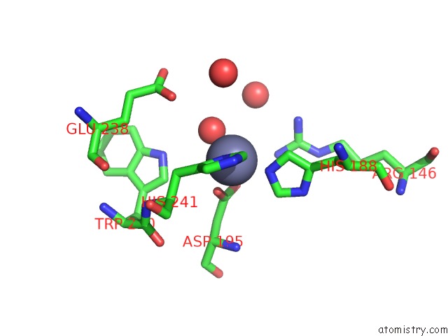
Mono view
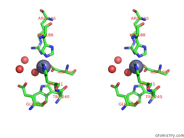
Stereo pair view
Mono view
Stereo pair view
A full contact list of Zinc with other atoms in the Zn binding
site number 1 of Structure of Vanyb Unbound within 5.0Å range:
|
Zinc binding site 2 out of 2 in 5zhf
Go back to
Zinc binding site 2 out
of 2 in the Structure of Vanyb Unbound
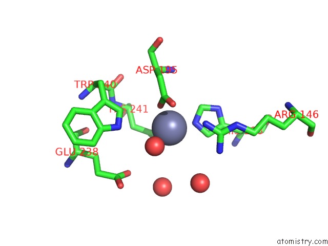
Mono view
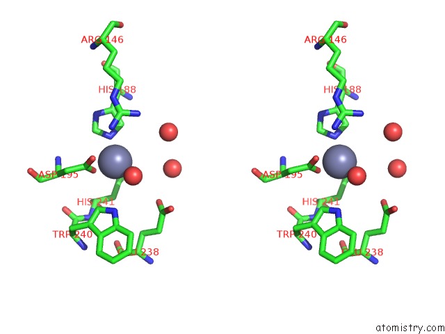
Stereo pair view
Mono view
Stereo pair view
A full contact list of Zinc with other atoms in the Zn binding
site number 2 of Structure of Vanyb Unbound within 5.0Å range:
|
Reference:
H.S.Kim,
H.Hahn,
J.Kim,
D.M.Jang,
J.Y.Lee,
J.M.Back,
H.N.Im,
H.Kim,
B.W.Han,
S.W.Suh.
Structural Basis For the Substrate Recognition of Peptidoglycan Pentapeptides By Enterococcus Faecalis Vanyb. Int. J. Biol. Macromol. V. 119 335 2018.
ISSN: ISSN 1879-0003
PubMed: 30016658
DOI: 10.1016/J.IJBIOMAC.2018.07.081
Page generated: Mon Oct 28 16:52:43 2024
ISSN: ISSN 1879-0003
PubMed: 30016658
DOI: 10.1016/J.IJBIOMAC.2018.07.081
Last articles
Zn in 9J0NZn in 9J0O
Zn in 9J0P
Zn in 9FJX
Zn in 9EKB
Zn in 9C0F
Zn in 9CAH
Zn in 9CH0
Zn in 9CH3
Zn in 9CH1