Zinc »
PDB 5yo1-5yy0 »
5yql »
Zinc in PDB 5yql: Crystal Structure of SIRT2 in Complex with Selective Inhibitor A2I
Protein crystallography data
The structure of Crystal Structure of SIRT2 in Complex with Selective Inhibitor A2I, PDB code: 5yql
was solved by
H.Wang,
Y.Yu,
G.Li,
Q.Chen,
with X-Ray Crystallography technique. A brief refinement statistics is given in the table below:
| Resolution Low / High (Å) | 35.90 / 1.60 |
| Space group | P 1 21 1 |
| Cell size a, b, c (Å), α, β, γ (°) | 36.038, 73.950, 55.797, 90.00, 95.11, 90.00 |
| R / Rfree (%) | 15.8 / 18.5 |
Zinc Binding Sites:
The binding sites of Zinc atom in the Crystal Structure of SIRT2 in Complex with Selective Inhibitor A2I
(pdb code 5yql). This binding sites where shown within
5.0 Angstroms radius around Zinc atom.
In total only one binding site of Zinc was determined in the Crystal Structure of SIRT2 in Complex with Selective Inhibitor A2I, PDB code: 5yql:
In total only one binding site of Zinc was determined in the Crystal Structure of SIRT2 in Complex with Selective Inhibitor A2I, PDB code: 5yql:
Zinc binding site 1 out of 1 in 5yql
Go back to
Zinc binding site 1 out
of 1 in the Crystal Structure of SIRT2 in Complex with Selective Inhibitor A2I
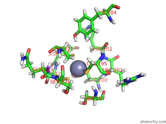
Mono view
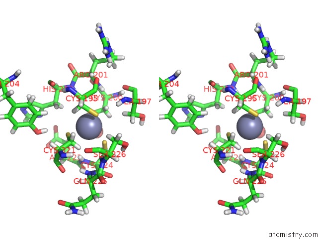
Stereo pair view
Mono view
Stereo pair view
A full contact list of Zinc with other atoms in the Zn binding
site number 1 of Crystal Structure of SIRT2 in Complex with Selective Inhibitor A2I within 5.0Å range:
|
Reference:
L.L.Yang,
H.L.Wang,
L.Zhong,
C.Yuan,
S.Y.Liu,
Z.J.Yu,
S.Liu,
Y.H.Yan,
C.Wu,
Y.Wang,
Z.Wang,
Y.Yu,
Q.Chen,
G.B.Li.
X-Ray Crystal Structure Guided Discovery of New Selective, Substrate-Mimicking Sirtuin 2 Inhibitors That Exhibit Activities Against Non-Small Cell Lung Cancer Cells. Eur J Med Chem V. 155 806 2018.
ISSN: ISSN 1768-3254
PubMed: 29957526
DOI: 10.1016/J.EJMECH.2018.06.041
Page generated: Mon Oct 28 16:16:32 2024
ISSN: ISSN 1768-3254
PubMed: 29957526
DOI: 10.1016/J.EJMECH.2018.06.041
Last articles
Zn in 9J0NZn in 9J0O
Zn in 9J0P
Zn in 9FJX
Zn in 9EKB
Zn in 9C0F
Zn in 9CAH
Zn in 9CH0
Zn in 9CH3
Zn in 9CH1