Zinc »
PDB 5yo1-5yy0 »
5yp8 »
Zinc in PDB 5yp8: P62/SQSTM1 Zz Domain with Arg-Peptide
Protein crystallography data
The structure of P62/SQSTM1 Zz Domain with Arg-Peptide, PDB code: 5yp8
was solved by
D.H.Kwon,
L.Kim,
H.K.Song,
with X-Ray Crystallography technique. A brief refinement statistics is given in the table below:
| Resolution Low / High (Å) | 19.81 / 1.45 |
| Space group | P 21 21 21 |
| Cell size a, b, c (Å), α, β, γ (°) | 28.981, 43.948, 66.596, 90.00, 90.00, 90.00 |
| R / Rfree (%) | 15.9 / 19.4 |
Zinc Binding Sites:
The binding sites of Zinc atom in the P62/SQSTM1 Zz Domain with Arg-Peptide
(pdb code 5yp8). This binding sites where shown within
5.0 Angstroms radius around Zinc atom.
In total 4 binding sites of Zinc where determined in the P62/SQSTM1 Zz Domain with Arg-Peptide, PDB code: 5yp8:
Jump to Zinc binding site number: 1; 2; 3; 4;
In total 4 binding sites of Zinc where determined in the P62/SQSTM1 Zz Domain with Arg-Peptide, PDB code: 5yp8:
Jump to Zinc binding site number: 1; 2; 3; 4;
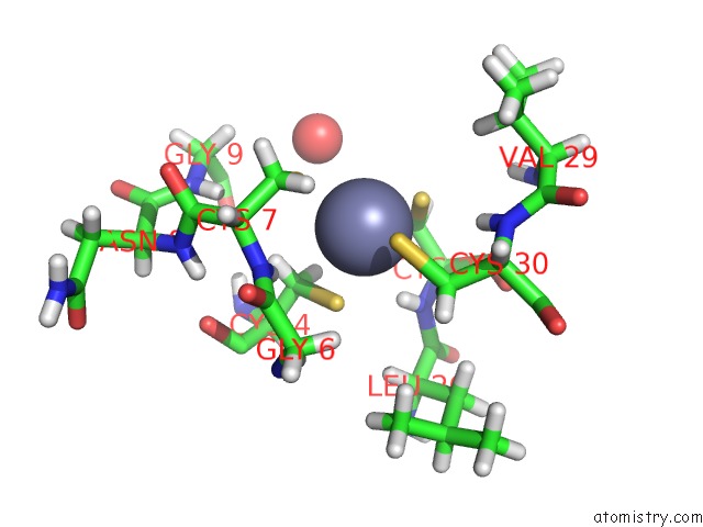
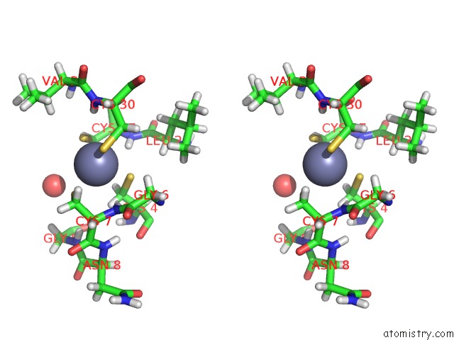
Zinc binding site 1 out of 4 in 5yp8
Go back to
Zinc binding site 1 out
of 4 in the P62/SQSTM1 Zz Domain with Arg-Peptide
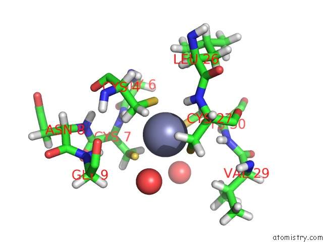
Mono view
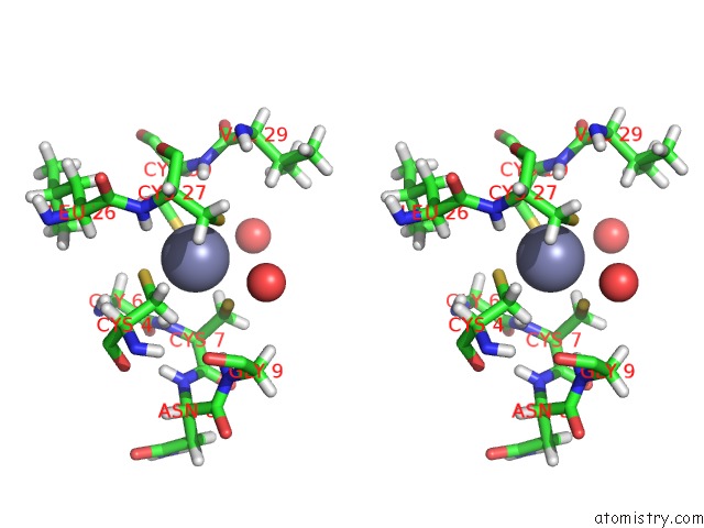
Stereo pair view
Mono view
Stereo pair view
A full contact list of Zinc with other atoms in the Zn binding
site number 1 of P62/SQSTM1 Zz Domain with Arg-Peptide within 5.0Å range:
|
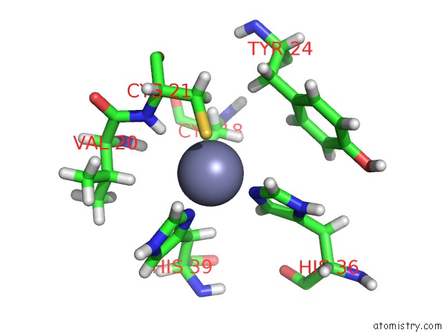
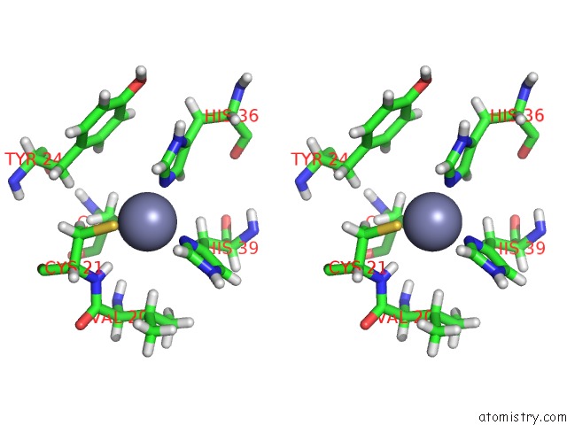
Zinc binding site 2 out of 4 in 5yp8
Go back to
Zinc binding site 2 out
of 4 in the P62/SQSTM1 Zz Domain with Arg-Peptide
Mono view
Stereo pair view
Mono view
Stereo pair view
A full contact list of Zinc with other atoms in the Zn binding
site number 2 of P62/SQSTM1 Zz Domain with Arg-Peptide within 5.0Å range:
|
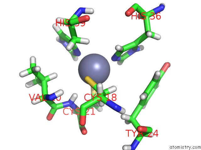
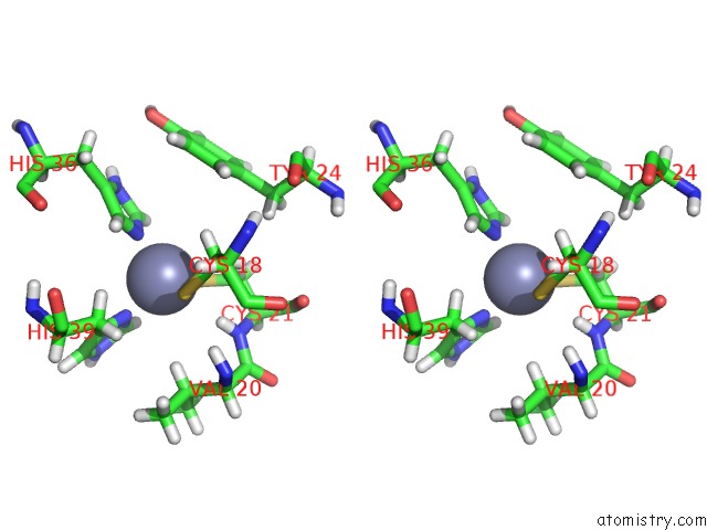
Zinc binding site 3 out of 4 in 5yp8
Go back to
Zinc binding site 3 out
of 4 in the P62/SQSTM1 Zz Domain with Arg-Peptide
Mono view
Stereo pair view
Mono view
Stereo pair view
A full contact list of Zinc with other atoms in the Zn binding
site number 3 of P62/SQSTM1 Zz Domain with Arg-Peptide within 5.0Å range:
|
Zinc binding site 4 out of 4 in 5yp8
Go back to
Zinc binding site 4 out
of 4 in the P62/SQSTM1 Zz Domain with Arg-Peptide
Mono view
Stereo pair view
Mono view
Stereo pair view
A full contact list of Zinc with other atoms in the Zn binding
site number 4 of P62/SQSTM1 Zz Domain with Arg-Peptide within 5.0Å range:
|
Reference:
D.H.Kwon,
O.H.Park,
L.Kim,
Y.O.Jung,
Y.Park,
H.Jeong,
J.Hyun,
Y.K.Kim,
H.K.Song.
Insights Into Degradation Mechanism of N-End Rule Substrates By P62/SQSTM1 Autophagy Adapter. Nat Commun V. 9 3291 2018.
ISSN: ESSN 2041-1723
PubMed: 30120248
DOI: 10.1038/S41467-018-05825-X
Page generated: Mon Oct 28 16:05:59 2024
ISSN: ESSN 2041-1723
PubMed: 30120248
DOI: 10.1038/S41467-018-05825-X
Last articles
Zn in 9J0NZn in 9J0O
Zn in 9J0P
Zn in 9FJX
Zn in 9EKB
Zn in 9C0F
Zn in 9CAH
Zn in 9CH0
Zn in 9CH3
Zn in 9CH1