Zinc »
PDB 5xob-5y1s »
5xob »
Zinc in PDB 5xob: Crystal Structure of Apo Tias (TRNAILE2 Agmatidine Synthetase)
Enzymatic activity of Crystal Structure of Apo Tias (TRNAILE2 Agmatidine Synthetase)
All present enzymatic activity of Crystal Structure of Apo Tias (TRNAILE2 Agmatidine Synthetase):
6.3.4.22;
6.3.4.22;
Protein crystallography data
The structure of Crystal Structure of Apo Tias (TRNAILE2 Agmatidine Synthetase), PDB code: 5xob
was solved by
J.Dong,
with X-Ray Crystallography technique. A brief refinement statistics is given in the table below:
| Resolution Low / High (Å) | 49.29 / 2.48 |
| Space group | P 41 21 2 |
| Cell size a, b, c (Å), α, β, γ (°) | 68.926, 68.926, 211.102, 90.00, 90.00, 90.00 |
| R / Rfree (%) | 24.5 / 29.4 |
Other elements in 5xob:
The structure of Crystal Structure of Apo Tias (TRNAILE2 Agmatidine Synthetase) also contains other interesting chemical elements:
| Magnesium | (Mg) | 1 atom |
Zinc Binding Sites:
The binding sites of Zinc atom in the Crystal Structure of Apo Tias (TRNAILE2 Agmatidine Synthetase)
(pdb code 5xob). This binding sites where shown within
5.0 Angstroms radius around Zinc atom.
In total only one binding site of Zinc was determined in the Crystal Structure of Apo Tias (TRNAILE2 Agmatidine Synthetase), PDB code: 5xob:
In total only one binding site of Zinc was determined in the Crystal Structure of Apo Tias (TRNAILE2 Agmatidine Synthetase), PDB code: 5xob:
Zinc binding site 1 out of 1 in 5xob
Go back to
Zinc binding site 1 out
of 1 in the Crystal Structure of Apo Tias (TRNAILE2 Agmatidine Synthetase)
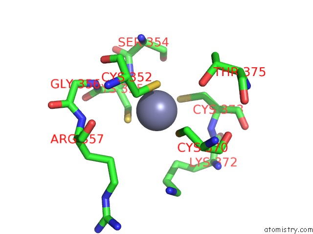
Mono view
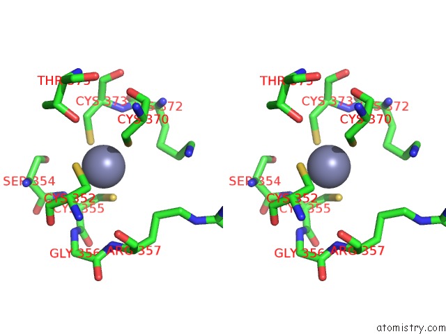
Stereo pair view
Mono view
Stereo pair view
A full contact list of Zinc with other atoms in the Zn binding
site number 1 of Crystal Structure of Apo Tias (TRNAILE2 Agmatidine Synthetase) within 5.0Å range:
|
Reference:
J.Dong,
F.Li,
F.Gao,
J.Wei,
Y.Lin,
Y.Zhang,
J.Lou,
G.Liu,
Y.Dong,
L.Liu,
H.Liu,
J.Wang,
W.Gong.
Structure of Trna-Modifying Enzyme Tias and Motions of Its Substrate Binding Zinc Ribbon. J. Mol. Biol. V. 430 4183 2018.
ISSN: ESSN 1089-8638
PubMed: 30121296
DOI: 10.1016/J.JMB.2018.08.015
Page generated: Mon Oct 28 15:11:23 2024
ISSN: ESSN 1089-8638
PubMed: 30121296
DOI: 10.1016/J.JMB.2018.08.015
Last articles
Zn in 9J0NZn in 9J0O
Zn in 9J0P
Zn in 9FJX
Zn in 9EKB
Zn in 9C0F
Zn in 9CAH
Zn in 9CH0
Zn in 9CH3
Zn in 9CH1