Zinc in PDB 5x6h: Crystal Structure of SMAD5-MH1/Gc-Bre Dna Complex
Protein crystallography data
The structure of Crystal Structure of SMAD5-MH1/Gc-Bre Dna Complex, PDB code: 5x6h
was solved by
N.Chai,
J.Wang,
Z.X.Wang,
J.W.Wu,
with X-Ray Crystallography technique. A brief refinement statistics is given in the table below:
| Resolution Low / High (Å) | 46.43 / 3.10 |
| Space group | P 64 2 2 |
| Cell size a, b, c (Å), α, β, γ (°) | 92.867, 92.867, 83.709, 90.00, 90.00, 120.00 |
| R / Rfree (%) | 20.2 / 23.1 |
Zinc Binding Sites:
The binding sites of Zinc atom in the Crystal Structure of SMAD5-MH1/Gc-Bre Dna Complex
(pdb code 5x6h). This binding sites where shown within
5.0 Angstroms radius around Zinc atom.
In total only one binding site of Zinc was determined in the Crystal Structure of SMAD5-MH1/Gc-Bre Dna Complex, PDB code: 5x6h:
In total only one binding site of Zinc was determined in the Crystal Structure of SMAD5-MH1/Gc-Bre Dna Complex, PDB code: 5x6h:
Zinc binding site 1 out of 1 in 5x6h
Go back to
Zinc binding site 1 out
of 1 in the Crystal Structure of SMAD5-MH1/Gc-Bre Dna Complex
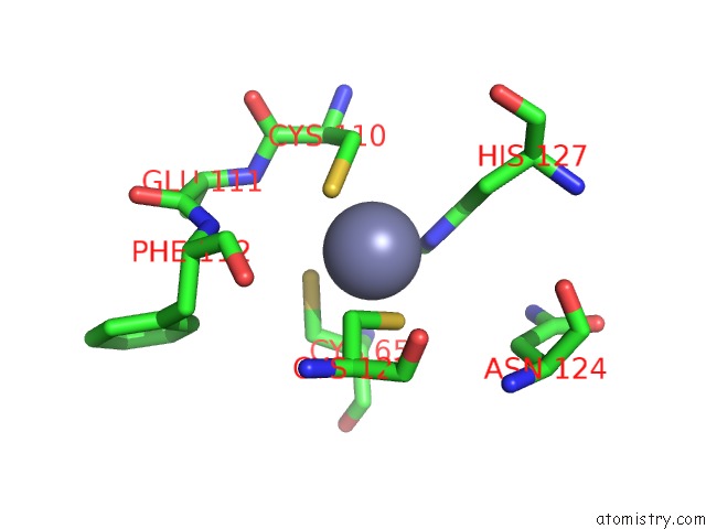
Mono view
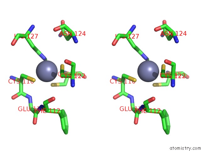
Stereo pair view
Mono view
Stereo pair view
A full contact list of Zinc with other atoms in the Zn binding
site number 1 of Crystal Structure of SMAD5-MH1/Gc-Bre Dna Complex within 5.0Å range:
|
Reference:
N.Chai,
W.X.Li,
J.Wang,
Z.X.Wang,
S.M.Yang,
J.W.Wu.
Structural Basis For the SMAD5 MH1 Domain to Recognize Different Dna Sequences. Nucleic Acids Res. V. 43 9051 2015.
ISSN: ESSN 1362-4962
PubMed: 26304548
DOI: 10.1093/NAR/GKV848
Page generated: Mon Oct 28 14:55:21 2024
ISSN: ESSN 1362-4962
PubMed: 26304548
DOI: 10.1093/NAR/GKV848
Last articles
Zn in 9MJ5Zn in 9HNW
Zn in 9G0L
Zn in 9FNE
Zn in 9DZN
Zn in 9E0I
Zn in 9D32
Zn in 9DAK
Zn in 8ZXC
Zn in 8ZUF