Zinc »
PDB 5wgw-5wpn »
5wob »
Zinc in PDB 5wob: Crystal Structure Analysis of FAB1-Bound Human Insulin Degrading Enzyme (Ide) in Complex with Insulin
Enzymatic activity of Crystal Structure Analysis of FAB1-Bound Human Insulin Degrading Enzyme (Ide) in Complex with Insulin
All present enzymatic activity of Crystal Structure Analysis of FAB1-Bound Human Insulin Degrading Enzyme (Ide) in Complex with Insulin:
3.4.24.56;
3.4.24.56;
Protein crystallography data
The structure of Crystal Structure Analysis of FAB1-Bound Human Insulin Degrading Enzyme (Ide) in Complex with Insulin, PDB code: 5wob
was solved by
L.A.Mccord,
W.G.Liang,
M.Farcasanu,
A.G.Wang,
S.Koide,
W.J.Tang,
with X-Ray Crystallography technique. A brief refinement statistics is given in the table below:
| Resolution Low / High (Å) | 49.54 / 3.95 |
| Space group | P 1 21 1 |
| Cell size a, b, c (Å), α, β, γ (°) | 121.589, 138.190, 376.509, 90.00, 99.36, 90.00 |
| R / Rfree (%) | 24.3 / 29.1 |
Zinc Binding Sites:
The binding sites of Zinc atom in the Crystal Structure Analysis of FAB1-Bound Human Insulin Degrading Enzyme (Ide) in Complex with Insulin
(pdb code 5wob). This binding sites where shown within
5.0 Angstroms radius around Zinc atom.
In total 8 binding sites of Zinc where determined in the Crystal Structure Analysis of FAB1-Bound Human Insulin Degrading Enzyme (Ide) in Complex with Insulin, PDB code: 5wob:
Jump to Zinc binding site number: 1; 2; 3; 4; 5; 6; 7; 8;
In total 8 binding sites of Zinc where determined in the Crystal Structure Analysis of FAB1-Bound Human Insulin Degrading Enzyme (Ide) in Complex with Insulin, PDB code: 5wob:
Jump to Zinc binding site number: 1; 2; 3; 4; 5; 6; 7; 8;
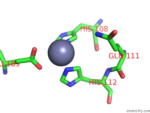
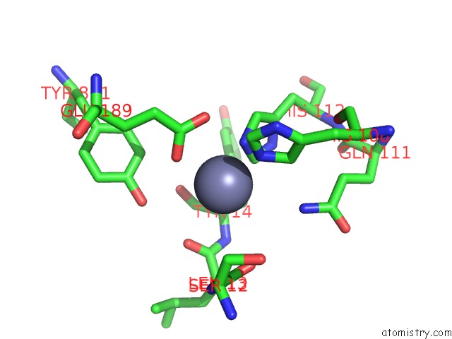
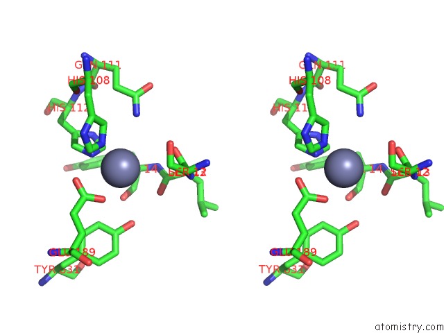
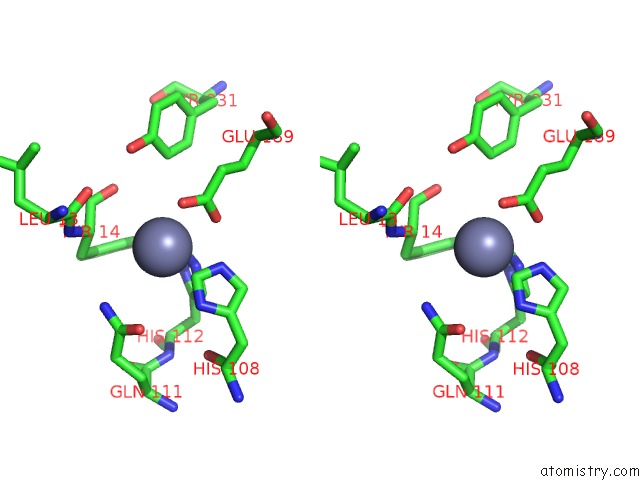
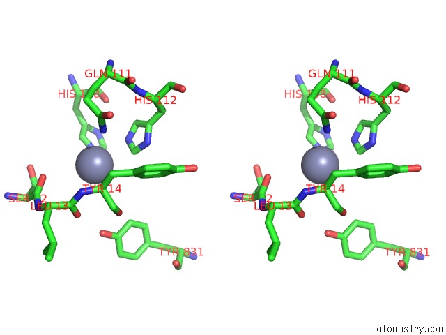
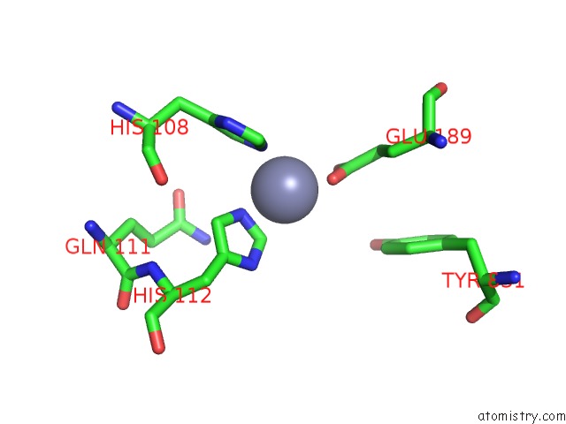
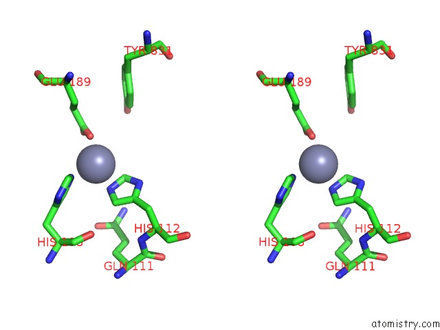
Zinc binding site 1 out of 8 in 5wob
Go back to
Zinc binding site 1 out
of 8 in the Crystal Structure Analysis of FAB1-Bound Human Insulin Degrading Enzyme (Ide) in Complex with Insulin
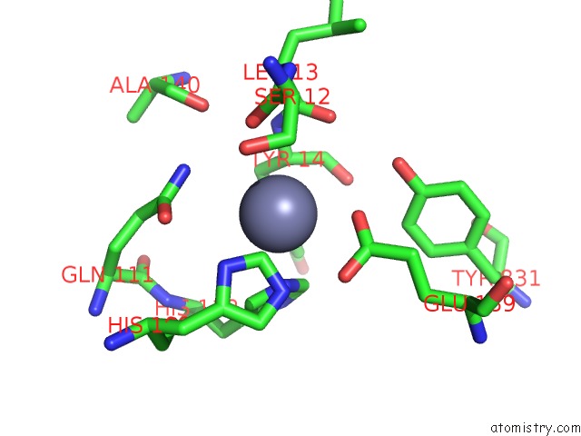
Mono view

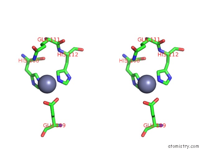
Stereo pair view
Mono view
Stereo pair view
A full contact list of Zinc with other atoms in the Zn binding
site number 1 of Crystal Structure Analysis of FAB1-Bound Human Insulin Degrading Enzyme (Ide) in Complex with Insulin within 5.0Å range:
|
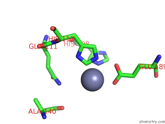
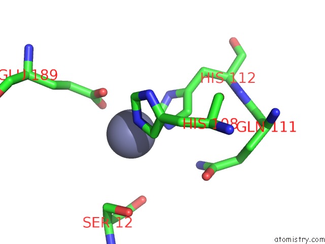
Zinc binding site 2 out of 8 in 5wob
Go back to
Zinc binding site 2 out
of 8 in the Crystal Structure Analysis of FAB1-Bound Human Insulin Degrading Enzyme (Ide) in Complex with Insulin
Mono view
Stereo pair view
Mono view
Stereo pair view
A full contact list of Zinc with other atoms in the Zn binding
site number 2 of Crystal Structure Analysis of FAB1-Bound Human Insulin Degrading Enzyme (Ide) in Complex with Insulin within 5.0Å range:
|
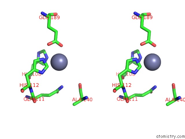
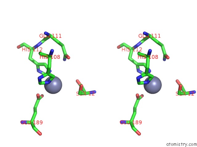
Zinc binding site 3 out of 8 in 5wob
Go back to
Zinc binding site 3 out
of 8 in the Crystal Structure Analysis of FAB1-Bound Human Insulin Degrading Enzyme (Ide) in Complex with Insulin
Mono view
Stereo pair view
Mono view
Stereo pair view
A full contact list of Zinc with other atoms in the Zn binding
site number 3 of Crystal Structure Analysis of FAB1-Bound Human Insulin Degrading Enzyme (Ide) in Complex with Insulin within 5.0Å range:
|
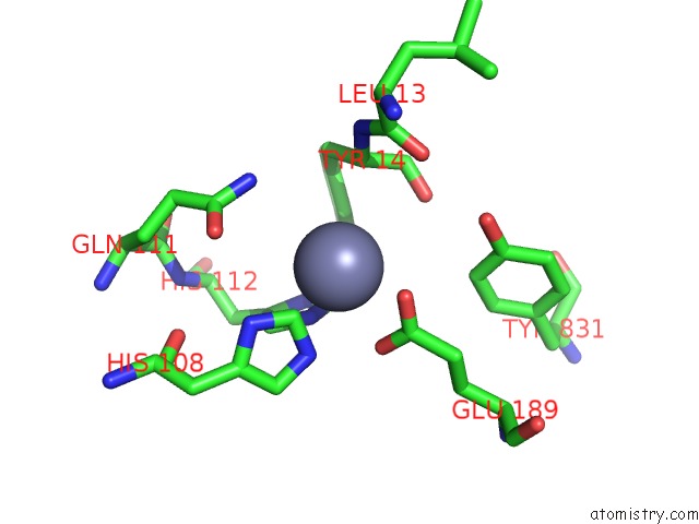
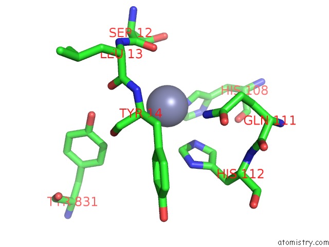
Zinc binding site 4 out of 8 in 5wob
Go back to
Zinc binding site 4 out
of 8 in the Crystal Structure Analysis of FAB1-Bound Human Insulin Degrading Enzyme (Ide) in Complex with Insulin
Mono view
Stereo pair view
Mono view
Stereo pair view
A full contact list of Zinc with other atoms in the Zn binding
site number 4 of Crystal Structure Analysis of FAB1-Bound Human Insulin Degrading Enzyme (Ide) in Complex with Insulin within 5.0Å range:
|
Zinc binding site 5 out of 8 in 5wob
Go back to
Zinc binding site 5 out
of 8 in the Crystal Structure Analysis of FAB1-Bound Human Insulin Degrading Enzyme (Ide) in Complex with Insulin
Mono view
Stereo pair view
Mono view
Stereo pair view
A full contact list of Zinc with other atoms in the Zn binding
site number 5 of Crystal Structure Analysis of FAB1-Bound Human Insulin Degrading Enzyme (Ide) in Complex with Insulin within 5.0Å range:
|
Zinc binding site 6 out of 8 in 5wob
Go back to
Zinc binding site 6 out
of 8 in the Crystal Structure Analysis of FAB1-Bound Human Insulin Degrading Enzyme (Ide) in Complex with Insulin
Mono view
Stereo pair view
Mono view
Stereo pair view
A full contact list of Zinc with other atoms in the Zn binding
site number 6 of Crystal Structure Analysis of FAB1-Bound Human Insulin Degrading Enzyme (Ide) in Complex with Insulin within 5.0Å range:
|
Zinc binding site 7 out of 8 in 5wob
Go back to
Zinc binding site 7 out
of 8 in the Crystal Structure Analysis of FAB1-Bound Human Insulin Degrading Enzyme (Ide) in Complex with Insulin
Mono view
Stereo pair view
Mono view
Stereo pair view
A full contact list of Zinc with other atoms in the Zn binding
site number 7 of Crystal Structure Analysis of FAB1-Bound Human Insulin Degrading Enzyme (Ide) in Complex with Insulin within 5.0Å range:
|
Zinc binding site 8 out of 8 in 5wob
Go back to
Zinc binding site 8 out
of 8 in the Crystal Structure Analysis of FAB1-Bound Human Insulin Degrading Enzyme (Ide) in Complex with Insulin
Mono view
Stereo pair view
Mono view
Stereo pair view
A full contact list of Zinc with other atoms in the Zn binding
site number 8 of Crystal Structure Analysis of FAB1-Bound Human Insulin Degrading Enzyme (Ide) in Complex with Insulin within 5.0Å range:
|
Reference:
Z.Zhang,
W.G.Liang,
L.J.Bailey,
Y.Z.Tan,
H.Wei,
A.Wang,
M.Farcasanu,
V.A.Woods,
L.A.Mccord,
D.Lee,
W.Shang,
R.Deprez-Poulain,
B.Deprez,
D.R.Liu,
A.Koide,
S.Koide,
A.A.Kossiakoff,
S.Li,
B.Carragher,
C.S.Potter,
W.J.Tang.
Ensemble Cryoem Elucidates the Mechanism of Insulin Capture and Degradation By Human Insulin Degrading Enzyme. Elife V. 7 2018.
ISSN: ESSN 2050-084X
PubMed: 29596046
DOI: 10.7554/ELIFE.33572
Page generated: Mon Oct 28 14:29:35 2024
ISSN: ESSN 2050-084X
PubMed: 29596046
DOI: 10.7554/ELIFE.33572
Last articles
Zn in 9J0NZn in 9J0O
Zn in 9J0P
Zn in 9FJX
Zn in 9EKB
Zn in 9C0F
Zn in 9CAH
Zn in 9CH0
Zn in 9CH3
Zn in 9CH1