Zinc »
PDB 5wgw-5wpn »
5wle »
Zinc in PDB 5wle: Crystal Structure of the Pps Phd Finger in Complex with H3K4ME3
Protein crystallography data
The structure of Crystal Structure of the Pps Phd Finger in Complex with H3K4ME3, PDB code: 5wle
was solved by
B.J.Klein,
T.G.Kutateladze,
with X-Ray Crystallography technique. A brief refinement statistics is given in the table below:
| Resolution Low / High (Å) | 31.20 / 1.95 |
| Space group | P 1 21 1 |
| Cell size a, b, c (Å), α, β, γ (°) | 22.972, 44.223, 31.308, 90.00, 94.77, 90.00 |
| R / Rfree (%) | 14.5 / 18.6 |
Zinc Binding Sites:
The binding sites of Zinc atom in the Crystal Structure of the Pps Phd Finger in Complex with H3K4ME3
(pdb code 5wle). This binding sites where shown within
5.0 Angstroms radius around Zinc atom.
In total 2 binding sites of Zinc where determined in the Crystal Structure of the Pps Phd Finger in Complex with H3K4ME3, PDB code: 5wle:
Jump to Zinc binding site number: 1; 2;
In total 2 binding sites of Zinc where determined in the Crystal Structure of the Pps Phd Finger in Complex with H3K4ME3, PDB code: 5wle:
Jump to Zinc binding site number: 1; 2;
Zinc binding site 1 out of 2 in 5wle
Go back to
Zinc binding site 1 out
of 2 in the Crystal Structure of the Pps Phd Finger in Complex with H3K4ME3
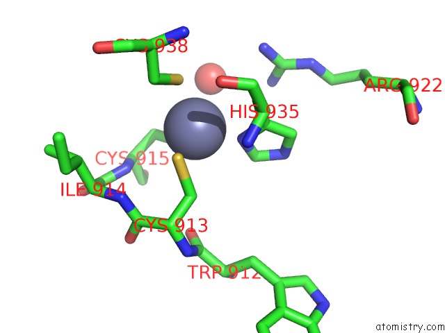
Mono view
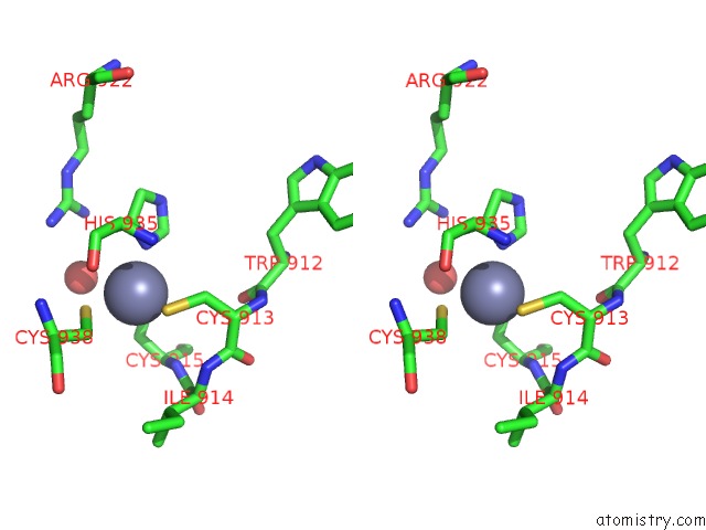
Stereo pair view
Mono view
Stereo pair view
A full contact list of Zinc with other atoms in the Zn binding
site number 1 of Crystal Structure of the Pps Phd Finger in Complex with H3K4ME3 within 5.0Å range:
|
Zinc binding site 2 out of 2 in 5wle
Go back to
Zinc binding site 2 out
of 2 in the Crystal Structure of the Pps Phd Finger in Complex with H3K4ME3
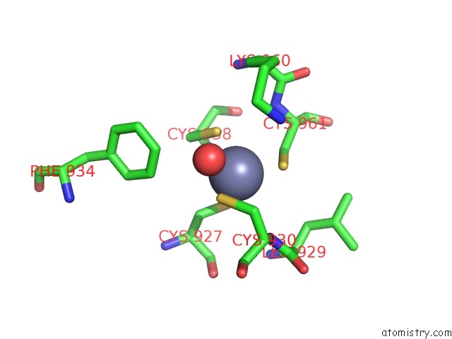
Mono view
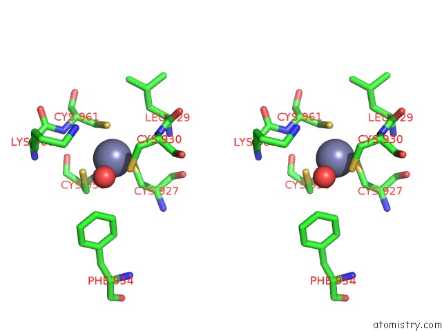
Stereo pair view
Mono view
Stereo pair view
A full contact list of Zinc with other atoms in the Zn binding
site number 2 of Crystal Structure of the Pps Phd Finger in Complex with H3K4ME3 within 5.0Å range:
|
Reference:
A.H.Tencer,
J.Gatchalian,
B.J.Klein,
A.Khan,
Y.Zhang,
B.D.Strahl,
K.H.M.Van Wely,
T.G.Kutateladze.
A Unique pH-Dependent Recognition of Methylated Histone H3K4 By Pps and Dido. Structure V. 25 1530 2017.
ISSN: ISSN 1878-4186
PubMed: 28919441
DOI: 10.1016/J.STR.2017.08.009
Page generated: Mon Oct 28 14:20:05 2024
ISSN: ISSN 1878-4186
PubMed: 28919441
DOI: 10.1016/J.STR.2017.08.009
Last articles
Zn in 9J0NZn in 9J0O
Zn in 9J0P
Zn in 9FJX
Zn in 9EKB
Zn in 9C0F
Zn in 9CAH
Zn in 9CH0
Zn in 9CH3
Zn in 9CH1