Zinc »
PDB 5wai-5wgw »
5wai »
Zinc in PDB 5wai: Crystal Structure of A SUZ12-RBBP4-JARID2-AEBP2 Heterotetrameric Complex
Protein crystallography data
The structure of Crystal Structure of A SUZ12-RBBP4-JARID2-AEBP2 Heterotetrameric Complex, PDB code: 5wai
was solved by
S.Chen,
L.Jiao,
X.Liu,
with X-Ray Crystallography technique. A brief refinement statistics is given in the table below:
| Resolution Low / High (Å) | 48.10 / 2.90 |
| Space group | P 21 21 21 |
| Cell size a, b, c (Å), α, β, γ (°) | 95.300, 111.428, 253.254, 90.00, 90.00, 90.00 |
| R / Rfree (%) | 17.1 / 21.6 |
Zinc Binding Sites:
The binding sites of Zinc atom in the Crystal Structure of A SUZ12-RBBP4-JARID2-AEBP2 Heterotetrameric Complex
(pdb code 5wai). This binding sites where shown within
5.0 Angstroms radius around Zinc atom.
In total 2 binding sites of Zinc where determined in the Crystal Structure of A SUZ12-RBBP4-JARID2-AEBP2 Heterotetrameric Complex, PDB code: 5wai:
Jump to Zinc binding site number: 1; 2;
In total 2 binding sites of Zinc where determined in the Crystal Structure of A SUZ12-RBBP4-JARID2-AEBP2 Heterotetrameric Complex, PDB code: 5wai:
Jump to Zinc binding site number: 1; 2;
Zinc binding site 1 out of 2 in 5wai
Go back to
Zinc binding site 1 out
of 2 in the Crystal Structure of A SUZ12-RBBP4-JARID2-AEBP2 Heterotetrameric Complex
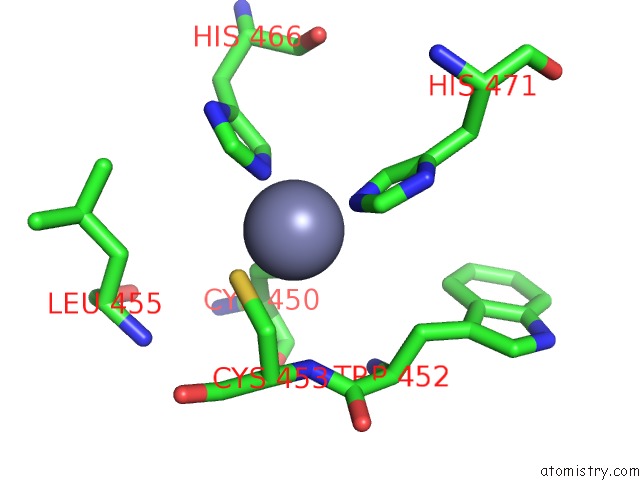
Mono view
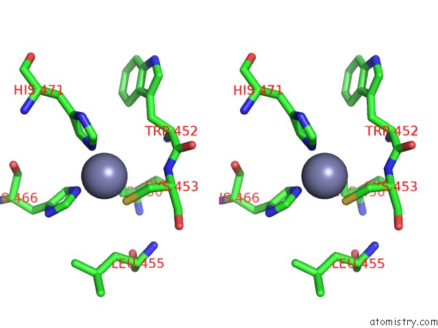
Stereo pair view
Mono view
Stereo pair view
A full contact list of Zinc with other atoms in the Zn binding
site number 1 of Crystal Structure of A SUZ12-RBBP4-JARID2-AEBP2 Heterotetrameric Complex within 5.0Å range:
|
Zinc binding site 2 out of 2 in 5wai
Go back to
Zinc binding site 2 out
of 2 in the Crystal Structure of A SUZ12-RBBP4-JARID2-AEBP2 Heterotetrameric Complex
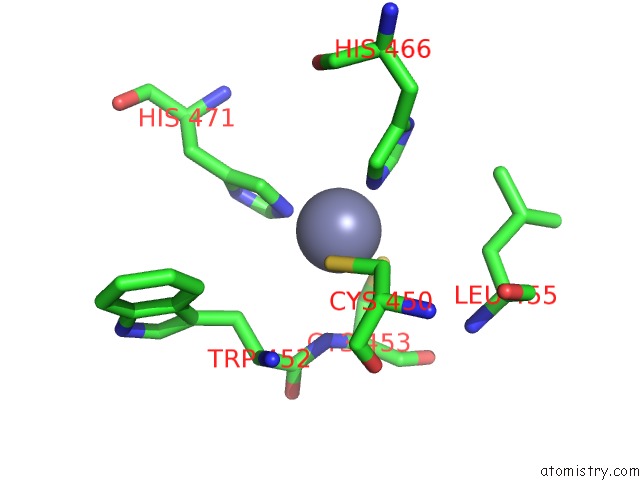
Mono view
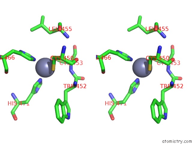
Stereo pair view
Mono view
Stereo pair view
A full contact list of Zinc with other atoms in the Zn binding
site number 2 of Crystal Structure of A SUZ12-RBBP4-JARID2-AEBP2 Heterotetrameric Complex within 5.0Å range:
|
Reference:
S.Chen,
L.Jiao,
M.Shubbar,
X.Yang,
X.Liu.
Unique Structural Platforms of SUZ12 Dictate Distinct Classes of PRC2 For Chromatin Binding. Mol. Cell V. 69 840 2018.
ISSN: ISSN 1097-4164
PubMed: 29499137
DOI: 10.1016/J.MOLCEL.2018.01.039
Page generated: Thu Aug 21 10:35:01 2025
ISSN: ISSN 1097-4164
PubMed: 29499137
DOI: 10.1016/J.MOLCEL.2018.01.039
Last articles
Zn in 6GYUZn in 6GYT
Zn in 6GYL
Zn in 6GYK
Zn in 6GXW
Zn in 6GXU
Zn in 6GXA
Zn in 6GXQ
Zn in 6GX3
Zn in 6GXE