Zinc »
PDB 5vmz-5vuq »
5vnn »
Zinc in PDB 5vnn: Crystal Structure of SEC23A/SEC24A/SEC22 Complexed with 4- Phenylbutyric Acid (50MM Soaking)
Protein crystallography data
The structure of Crystal Structure of SEC23A/SEC24A/SEC22 Complexed with 4- Phenylbutyric Acid (50MM Soaking), PDB code: 5vnn
was solved by
W.Ma,
J.Goldberg,
with X-Ray Crystallography technique. A brief refinement statistics is given in the table below:
| Resolution Low / High (Å) | 48.86 / 2.50 |
| Space group | C 1 2 1 |
| Cell size a, b, c (Å), α, β, γ (°) | 148.016, 97.715, 128.967, 90.00, 89.59, 90.00 |
| R / Rfree (%) | 21 / 25.1 |
Zinc Binding Sites:
The binding sites of Zinc atom in the Crystal Structure of SEC23A/SEC24A/SEC22 Complexed with 4- Phenylbutyric Acid (50MM Soaking)
(pdb code 5vnn). This binding sites where shown within
5.0 Angstroms radius around Zinc atom.
In total 2 binding sites of Zinc where determined in the Crystal Structure of SEC23A/SEC24A/SEC22 Complexed with 4- Phenylbutyric Acid (50MM Soaking), PDB code: 5vnn:
Jump to Zinc binding site number: 1; 2;
In total 2 binding sites of Zinc where determined in the Crystal Structure of SEC23A/SEC24A/SEC22 Complexed with 4- Phenylbutyric Acid (50MM Soaking), PDB code: 5vnn:
Jump to Zinc binding site number: 1; 2;
Zinc binding site 1 out of 2 in 5vnn
Go back to
Zinc binding site 1 out
of 2 in the Crystal Structure of SEC23A/SEC24A/SEC22 Complexed with 4- Phenylbutyric Acid (50MM Soaking)
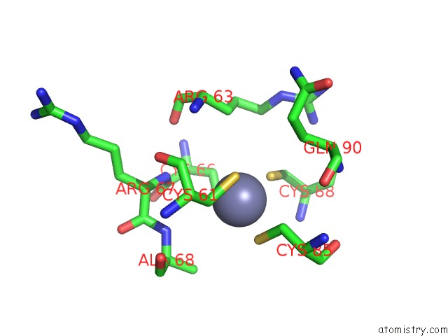
Mono view
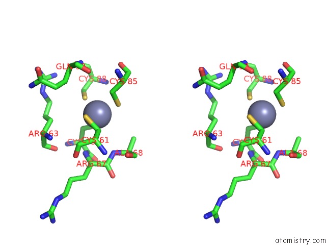
Stereo pair view
Mono view
Stereo pair view
A full contact list of Zinc with other atoms in the Zn binding
site number 1 of Crystal Structure of SEC23A/SEC24A/SEC22 Complexed with 4- Phenylbutyric Acid (50MM Soaking) within 5.0Å range:
|
Zinc binding site 2 out of 2 in 5vnn
Go back to
Zinc binding site 2 out
of 2 in the Crystal Structure of SEC23A/SEC24A/SEC22 Complexed with 4- Phenylbutyric Acid (50MM Soaking)

Mono view
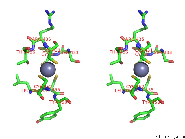
Stereo pair view
Mono view
Stereo pair view
A full contact list of Zinc with other atoms in the Zn binding
site number 2 of Crystal Structure of SEC23A/SEC24A/SEC22 Complexed with 4- Phenylbutyric Acid (50MM Soaking) within 5.0Å range:
|
Reference:
W.Ma,
E.Goldberg,
J.Goldberg.
Er Retention Is Imposed By Copii Protein Sorting and Attenuated By 4-Phenylbutyrate. Elife V. 6 2017.
ISSN: ESSN 2050-084X
PubMed: 28594326
DOI: 10.7554/ELIFE.26624
Page generated: Mon Oct 28 13:15:58 2024
ISSN: ESSN 2050-084X
PubMed: 28594326
DOI: 10.7554/ELIFE.26624
Last articles
Zn in 9J0NZn in 9J0O
Zn in 9J0P
Zn in 9FJX
Zn in 9EKB
Zn in 9C0F
Zn in 9CAH
Zn in 9CH0
Zn in 9CH3
Zn in 9CH1