Zinc »
PDB 5thi-5tt8 »
5tkq »
Zinc in PDB 5tkq: Crystal Structure of Human 3HAO with Zinc Bound in the Active Site
Enzymatic activity of Crystal Structure of Human 3HAO with Zinc Bound in the Active Site
All present enzymatic activity of Crystal Structure of Human 3HAO with Zinc Bound in the Active Site:
1.13.11.6;
1.13.11.6;
Protein crystallography data
The structure of Crystal Structure of Human 3HAO with Zinc Bound in the Active Site, PDB code: 5tkq
was solved by
L.S.Pidugu,
E.A.Toth,
with X-Ray Crystallography technique. A brief refinement statistics is given in the table below:
| Resolution Low / High (Å) | 38.15 / 1.75 |
| Space group | P 65 2 2 |
| Cell size a, b, c (Å), α, β, γ (°) | 57.336, 57.336, 417.100, 90.00, 90.00, 120.00 |
| R / Rfree (%) | 20.2 / 22.4 |
Zinc Binding Sites:
The binding sites of Zinc atom in the Crystal Structure of Human 3HAO with Zinc Bound in the Active Site
(pdb code 5tkq). This binding sites where shown within
5.0 Angstroms radius around Zinc atom.
In total only one binding site of Zinc was determined in the Crystal Structure of Human 3HAO with Zinc Bound in the Active Site, PDB code: 5tkq:
In total only one binding site of Zinc was determined in the Crystal Structure of Human 3HAO with Zinc Bound in the Active Site, PDB code: 5tkq:
Zinc binding site 1 out of 1 in 5tkq
Go back to
Zinc binding site 1 out
of 1 in the Crystal Structure of Human 3HAO with Zinc Bound in the Active Site
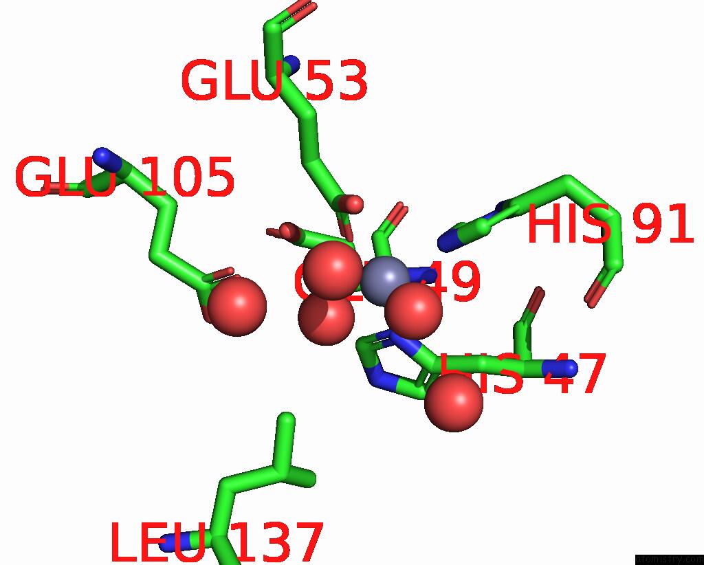
Mono view
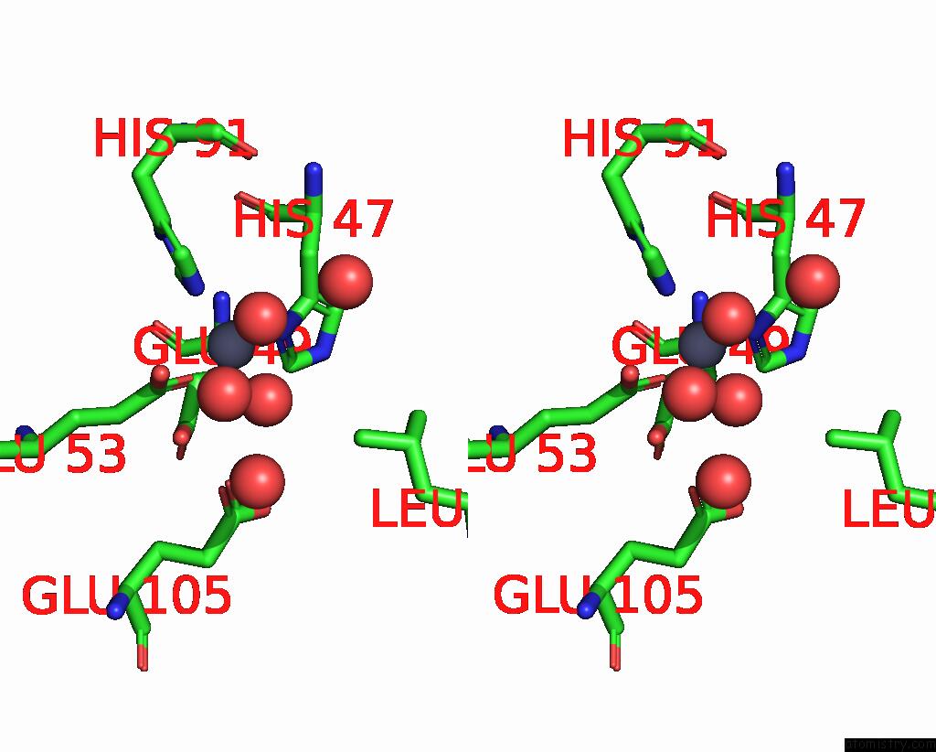
Stereo pair view
Mono view
Stereo pair view
A full contact list of Zinc with other atoms in the Zn binding
site number 1 of Crystal Structure of Human 3HAO with Zinc Bound in the Active Site within 5.0Å range:
|
Reference:
L.S.Pidugu,
H.Neu,
T.L.Wong,
E.Pozharski,
J.L.Molloy,
S.L.Michel,
E.A.Toth.
Crystal Structures of Human 3-Hydroxyanthranilate 3,4-Dioxygenase with Native and Non-Native Metals Bound in the Active Site. Acta Crystallogr D Struct V. 73 340 2017BIOL.
ISSN: ISSN 2059-7983
PubMed: 28375145
DOI: 10.1107/S2059798317002029
Page generated: Mon Oct 28 08:38:14 2024
ISSN: ISSN 2059-7983
PubMed: 28375145
DOI: 10.1107/S2059798317002029
Last articles
Zn in 9J0NZn in 9J0O
Zn in 9J0P
Zn in 9FJX
Zn in 9EKB
Zn in 9C0F
Zn in 9CAH
Zn in 9CH0
Zn in 9CH3
Zn in 9CH1