Zinc »
PDB 5pyu-5qpr »
5pz0 »
Zinc in PDB 5pz0: Pandda Analysis Group Deposition -- Crystal Structure of SP100 After Initial Refinement with No Ligand Modelled (Structure 96)
Protein crystallography data
The structure of Pandda Analysis Group Deposition -- Crystal Structure of SP100 After Initial Refinement with No Ligand Modelled (Structure 96), PDB code: 5pz0
was solved by
N.M.Pearce,
T.Krojer,
R.Talon,
A.R.Bradley,
M.Fairhead,
R.Sethi,
N.Wright,
E.Maclean,
P.Collins,
J.Brandao-Neto,
A.Douangamath,
Z.Renjie,
A.Dias,
J.Ng,
P.E.Brennan,
O.Cox,
C.Bountra,
C.H.Arrowsmith,
A.Edwards,
F.Vondelft,
with X-Ray Crystallography technique. A brief refinement statistics is given in the table below:
| Resolution Low / High (Å) | 39.05 / 2.13 |
| Space group | C 1 2 1 |
| Cell size a, b, c (Å), α, β, γ (°) | 127.720, 45.510, 83.630, 90.00, 101.90, 90.00 |
| R / Rfree (%) | 16.4 / 21.3 |
Zinc Binding Sites:
The binding sites of Zinc atom in the Pandda Analysis Group Deposition -- Crystal Structure of SP100 After Initial Refinement with No Ligand Modelled (Structure 96)
(pdb code 5pz0). This binding sites where shown within
5.0 Angstroms radius around Zinc atom.
In total 5 binding sites of Zinc where determined in the Pandda Analysis Group Deposition -- Crystal Structure of SP100 After Initial Refinement with No Ligand Modelled (Structure 96), PDB code: 5pz0:
Jump to Zinc binding site number: 1; 2; 3; 4; 5;
In total 5 binding sites of Zinc where determined in the Pandda Analysis Group Deposition -- Crystal Structure of SP100 After Initial Refinement with No Ligand Modelled (Structure 96), PDB code: 5pz0:
Jump to Zinc binding site number: 1; 2; 3; 4; 5;
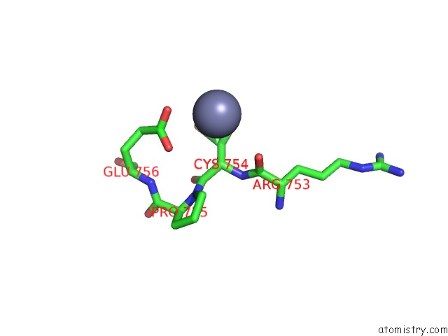
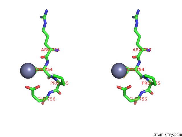
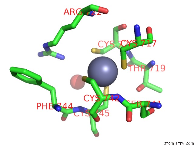
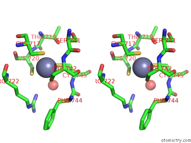
Zinc binding site 1 out of 5 in 5pz0
Go back to
Zinc binding site 1 out
of 5 in the Pandda Analysis Group Deposition -- Crystal Structure of SP100 After Initial Refinement with No Ligand Modelled (Structure 96)
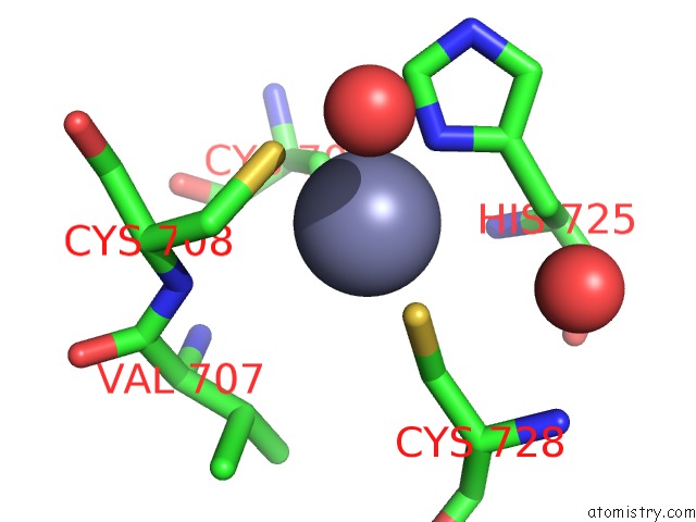
Mono view
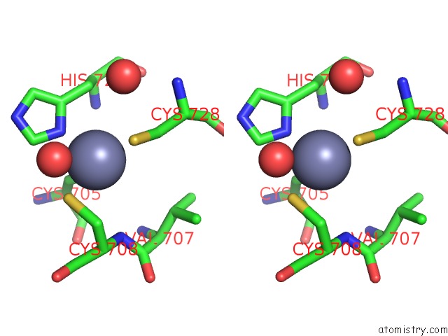
Stereo pair view
Mono view
Stereo pair view
A full contact list of Zinc with other atoms in the Zn binding
site number 1 of Pandda Analysis Group Deposition -- Crystal Structure of SP100 After Initial Refinement with No Ligand Modelled (Structure 96) within 5.0Å range:
|
Zinc binding site 2 out of 5 in 5pz0
Go back to
Zinc binding site 2 out
of 5 in the Pandda Analysis Group Deposition -- Crystal Structure of SP100 After Initial Refinement with No Ligand Modelled (Structure 96)
Mono view
Stereo pair view
Mono view
Stereo pair view
A full contact list of Zinc with other atoms in the Zn binding
site number 2 of Pandda Analysis Group Deposition -- Crystal Structure of SP100 After Initial Refinement with No Ligand Modelled (Structure 96) within 5.0Å range:
|
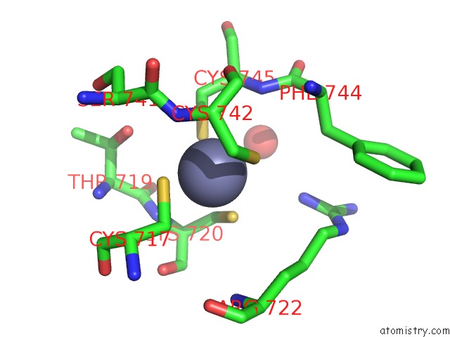
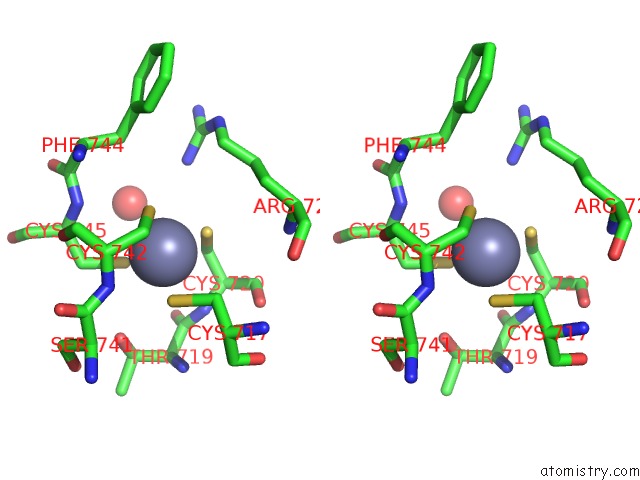
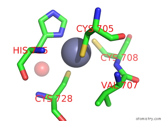
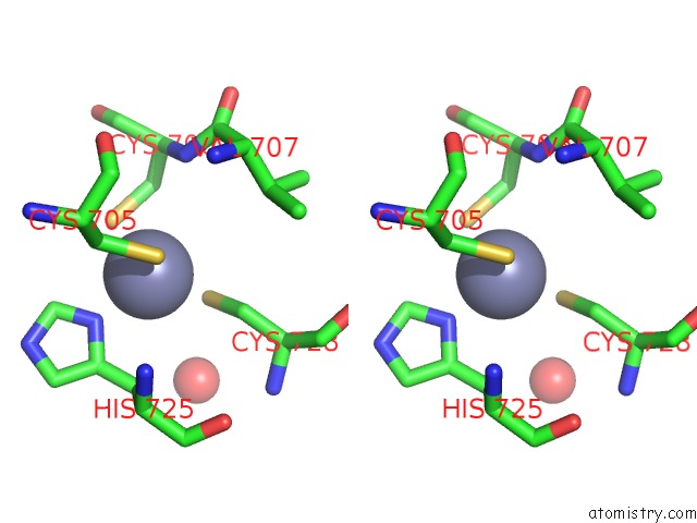
Zinc binding site 3 out of 5 in 5pz0
Go back to
Zinc binding site 3 out
of 5 in the Pandda Analysis Group Deposition -- Crystal Structure of SP100 After Initial Refinement with No Ligand Modelled (Structure 96)
Mono view
Stereo pair view
Mono view
Stereo pair view
A full contact list of Zinc with other atoms in the Zn binding
site number 3 of Pandda Analysis Group Deposition -- Crystal Structure of SP100 After Initial Refinement with No Ligand Modelled (Structure 96) within 5.0Å range:
|
Zinc binding site 4 out of 5 in 5pz0
Go back to
Zinc binding site 4 out
of 5 in the Pandda Analysis Group Deposition -- Crystal Structure of SP100 After Initial Refinement with No Ligand Modelled (Structure 96)
Mono view
Stereo pair view
Mono view
Stereo pair view
A full contact list of Zinc with other atoms in the Zn binding
site number 4 of Pandda Analysis Group Deposition -- Crystal Structure of SP100 After Initial Refinement with No Ligand Modelled (Structure 96) within 5.0Å range:
|
Zinc binding site 5 out of 5 in 5pz0
Go back to
Zinc binding site 5 out
of 5 in the Pandda Analysis Group Deposition -- Crystal Structure of SP100 After Initial Refinement with No Ligand Modelled (Structure 96)
Mono view
Stereo pair view
Mono view
Stereo pair view
A full contact list of Zinc with other atoms in the Zn binding
site number 5 of Pandda Analysis Group Deposition -- Crystal Structure of SP100 After Initial Refinement with No Ligand Modelled (Structure 96) within 5.0Å range:
|
Reference:
N.M.Pearce,
T.Krojer,
A.R.Bradley,
P.Collins,
R.P.Nowak,
R.Talon,
B.D.Marsden,
S.Kelm,
J.Shi,
C.M.Deane,
F.Von Delft.
A Multi-Crystal Method For Extracting Obscured Crystallographic States From Conventionally Uninterpretable Electron Density. Nat Commun V. 8 15123 2017.
ISSN: ESSN 2041-1723
PubMed: 28436492
DOI: 10.1038/NCOMMS15123
Page generated: Mon Oct 28 03:32:25 2024
ISSN: ESSN 2041-1723
PubMed: 28436492
DOI: 10.1038/NCOMMS15123
Last articles
Zn in 9J0NZn in 9J0O
Zn in 9J0P
Zn in 9FJX
Zn in 9EKB
Zn in 9C0F
Zn in 9CAH
Zn in 9CH0
Zn in 9CH3
Zn in 9CH1