Zinc »
PDB 5ofn-5onj »
5om9 »
Zinc in PDB 5om9: Crystal Structure of the Human Carboxypeptidase A1 in Complex with A Thiirane Mechanism-Based Inhibitor
Enzymatic activity of Crystal Structure of the Human Carboxypeptidase A1 in Complex with A Thiirane Mechanism-Based Inhibitor
All present enzymatic activity of Crystal Structure of the Human Carboxypeptidase A1 in Complex with A Thiirane Mechanism-Based Inhibitor:
3.4.17.1;
3.4.17.1;
Protein crystallography data
The structure of Crystal Structure of the Human Carboxypeptidase A1 in Complex with A Thiirane Mechanism-Based Inhibitor, PDB code: 5om9
was solved by
P.Gallego,
C.Granados,
D.Fernandez,
I.Pallares,
G.Covaleda,
F.X.Aviles,
J.Vendrell,
D.Reverter,
with X-Ray Crystallography technique. A brief refinement statistics is given in the table below:
| Resolution Low / High (Å) | 43.68 / 1.80 |
| Space group | P 1 21 1 |
| Cell size a, b, c (Å), α, β, γ (°) | 53.901, 52.544, 133.984, 90.00, 94.84, 90.00 |
| R / Rfree (%) | 15.4 / 21.2 |
Zinc Binding Sites:
The binding sites of Zinc atom in the Crystal Structure of the Human Carboxypeptidase A1 in Complex with A Thiirane Mechanism-Based Inhibitor
(pdb code 5om9). This binding sites where shown within
5.0 Angstroms radius around Zinc atom.
In total 3 binding sites of Zinc where determined in the Crystal Structure of the Human Carboxypeptidase A1 in Complex with A Thiirane Mechanism-Based Inhibitor, PDB code: 5om9:
Jump to Zinc binding site number: 1; 2; 3;
In total 3 binding sites of Zinc where determined in the Crystal Structure of the Human Carboxypeptidase A1 in Complex with A Thiirane Mechanism-Based Inhibitor, PDB code: 5om9:
Jump to Zinc binding site number: 1; 2; 3;
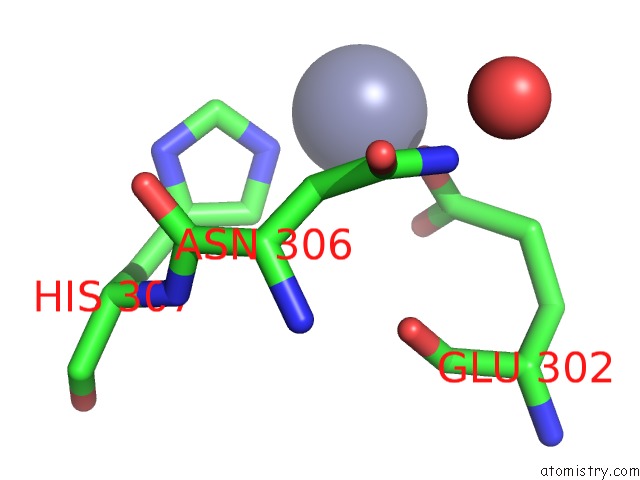
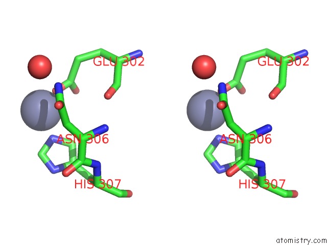
Zinc binding site 1 out of 3 in 5om9
Go back to
Zinc binding site 1 out
of 3 in the Crystal Structure of the Human Carboxypeptidase A1 in Complex with A Thiirane Mechanism-Based Inhibitor
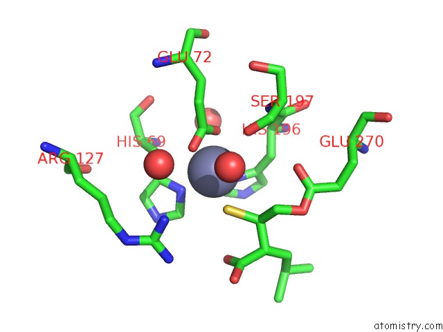
Mono view
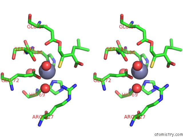
Stereo pair view
Mono view
Stereo pair view
A full contact list of Zinc with other atoms in the Zn binding
site number 1 of Crystal Structure of the Human Carboxypeptidase A1 in Complex with A Thiirane Mechanism-Based Inhibitor within 5.0Å range:
|
Zinc binding site 2 out of 3 in 5om9
Go back to
Zinc binding site 2 out
of 3 in the Crystal Structure of the Human Carboxypeptidase A1 in Complex with A Thiirane Mechanism-Based Inhibitor
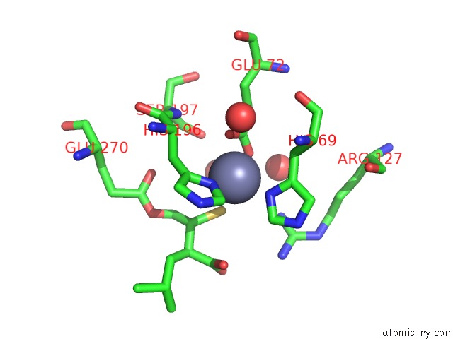
Mono view
Stereo pair view
Mono view
Stereo pair view
A full contact list of Zinc with other atoms in the Zn binding
site number 2 of Crystal Structure of the Human Carboxypeptidase A1 in Complex with A Thiirane Mechanism-Based Inhibitor within 5.0Å range:
|
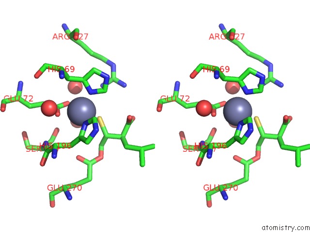
Zinc binding site 3 out of 3 in 5om9
Go back to
Zinc binding site 3 out
of 3 in the Crystal Structure of the Human Carboxypeptidase A1 in Complex with A Thiirane Mechanism-Based Inhibitor
Mono view
Stereo pair view
Mono view
Stereo pair view
A full contact list of Zinc with other atoms in the Zn binding
site number 3 of Crystal Structure of the Human Carboxypeptidase A1 in Complex with A Thiirane Mechanism-Based Inhibitor within 5.0Å range:
|
Reference:
S.A.Testero,
C.Granados,
D.Fernandez,
P.Gallego,
G.Covaleda,
D.Reverter,
J.Vendrell,
F.X.Aviles,
I.Pallares,
S.Mobashery.
Discovery of Mechanism-Based Inactivators For Human Pancreatic Carboxypeptidase A From A Focused Synthetic Library. Acs Med Chem Lett V. 8 1122 2017.
ISSN: ISSN 1948-5875
PubMed: 29057062
DOI: 10.1021/ACSMEDCHEMLETT.7B00346
Page generated: Sun Oct 27 23:41:09 2024
ISSN: ISSN 1948-5875
PubMed: 29057062
DOI: 10.1021/ACSMEDCHEMLETT.7B00346
Last articles
Zn in 9J0NZn in 9J0O
Zn in 9J0P
Zn in 9FJX
Zn in 9EKB
Zn in 9C0F
Zn in 9CAH
Zn in 9CH0
Zn in 9CH3
Zn in 9CH1